TLDR;
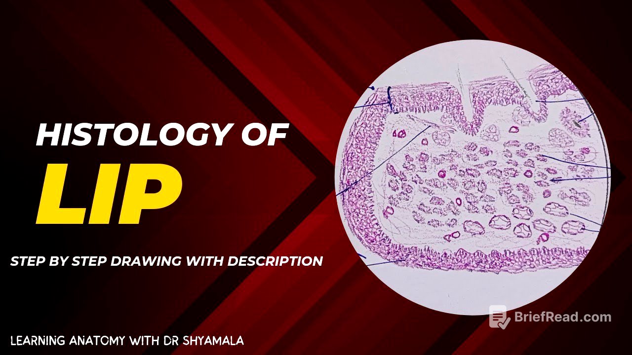
This video provides a detailed explanation of the histology of the lip, focusing on its three main regions: the outer surface (thin skin), the inner mucosal surface, and the transition zone (vermilion zone). It describes the types of epithelium found in each region, the presence of various glands and structures like hair follicles and muscles, and the importance of blood supply.
- The lip consists of three layers: outer skin, transition zone, and inner mucosal surface.
- The main bulk of the lip is the orbicularis oris muscle.
- Each layer has distinct histological features, including different types of epithelium and the presence of glands.
Introduction [0:00]
The video introduces a discussion on the histology of the lip, emphasizing its mobile nature due to a well-developed core of striated muscles. The lip is divided into three main regions: the outer surface, the inner mucosal surface, and the transition border.
Outer Surface (Thin Skin) [0:11]
The outer surface of the lip is composed of thin skin, characterized by stratified squamous keratinized epithelium. This layer includes hair follicles, arrector pili muscles, and sebaceous glands. The thickness of the hair follicles varies, with thicker follicles present near the mustache region of the upper lip and thinner follicles in the lower lip. The epithelium consists of basal columnar cells with vertically oval nuclei, polygonal cells with round nuclei, and flattened squamous cells with a thin layer of keratin.
Transition Zone (Vermilion Zone) [4:15]
The transition zone, also known as the Vermilion Zone, is covered by stratified squamous non-keratinized epithelium, although some sources suggest a very thin layer of keratin may be present. This zone is notable for its rich blood supply, which gives the lip its red color due to the presence of numerous capillaries and nerves. The basement membrane in this region is wavy, forming rete ridges that aid in the firm attachment of the epithelium to the underlying lamina propria.
Inner Mucosal Surface [5:20]
The inner mucosal surface is lined by stratified squamous non-keratinized epithelium. The basement membrane is folded, creating rete ridges. Underlying this layer are numerous mucosal and serosal glands that keep the surface moist. Unlike the transition zone, which is kept moist by saliva in the oral cavity, the mucosal surface has its own glands for moisture.
Orbicularis Oris Muscle [6:05]
The main bulk of the lip is composed of the orbicularis oris muscle, a skeletal muscle. Depending on the section, it can appear as a transverse or oblique section. The muscle is rich in capillaries, especially below the transition zone. Histologically, it consists of polygonal-shaped muscle fascicles with peripherally placed multiple nuclei, characteristic of skeletal muscle.
Conclusion [7:11]
The video concludes by summarizing the histology of the lip, highlighting the outer thin skin, inner mucosal surface, transition zone, and the main bulk of skeletal muscle. It emphasizes the presence of mucous glands in the mucosal surface for moisture and the hair and its apparatus in the thin skin. The video encourages viewers to refer to the labeled diagram provided at the end for further clarification.