TLDR;
The endoplasmic reticulum (ER) is a vast and dynamic organelle responsible for protein and lipid synthesis, calcium signaling, and glycosylation. Key points include:
- ER constitutes a significant portion of the cell's membranes and volume.
- It was discovered in 1954 by Keith Porter and George Palade using electron microscopy.
- ER membranes are crucial for protein synthesis and intracellular transport.
- Microsomes, formed from disrupted ER membranes, retain ER functions.
- ER morphology is linked to various pathological conditions.
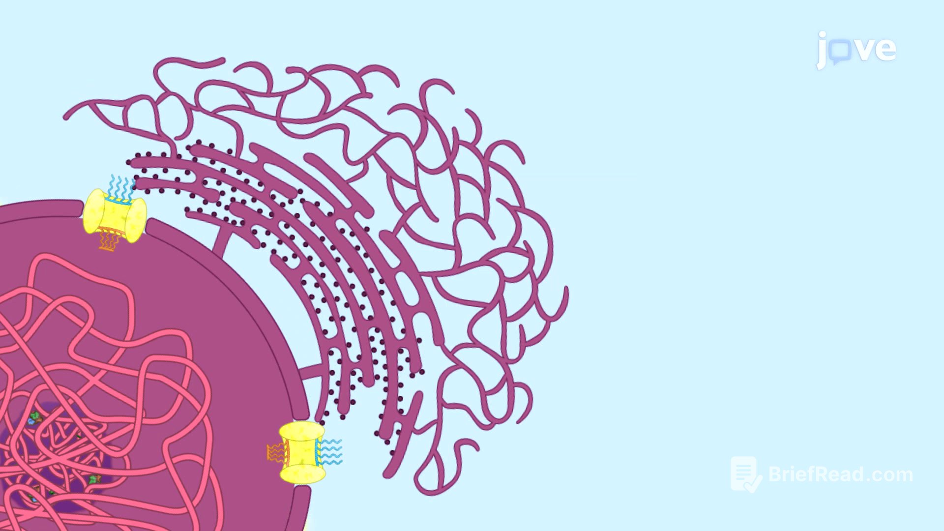
The Endoplasmic Reticulum
The endoplasmic reticulum (ER) comprises over half of a cell's membranes and 10% of its volume, serving as the primary site for protein and lipid synthesis for organelles like the Golgi apparatus and plasma membrane. Discovered in 1954 by Keith Porter and George Palade, the ER's membranes are essential for protein synthesis and intracellular transport, identified through radio-labeled and fluorescent-labeled amino acids. Isolation of the ER is challenging due to its intricate structure, but cell homogenization results in microsomes, small vesicles capable of sustaining ER functions such as protein and lipid synthesis, calcium signaling, and glycosylation. Subcellular fractionation, particularly using a sucrose gradient, is commonly used to purify these membranes, with rough ER microsomes sedimenting at a higher density than smooth ER microsomes. The ER network is dynamic, constantly changing shape with the cytoskeleton to provide mechanical support. Interconversion between cisternae and tubule morphologies is possible, regulated by membrane protein expression. Disruption of ER morphology is associated with diseases like Alzheimer's, hereditary spastic paraplegia, and viral infections such as hepatitis C and dengue.