TLDR;
This text describes Magnetic Resonance Imaging (MRI), a noninvasive medical imaging technique that utilizes magnetic fields and radio waves to create detailed images of the body's internal structures. It highlights the advantages of MRI, such as the absence of radiation exposure, and its drawbacks, including high costs and patient discomfort. The text also explains different types of MRI images, the use of contrast media, and advanced applications like functional MRI and 4D flow MRI.
- MRI is a noninvasive imaging technique using magnetic fields and radio waves.
- It offers detailed imaging without radiation exposure.
- Functional MRI and 4D flow MRI are advanced applications for studying brain activity and blood flow.
Magnetic Resonance Imaging
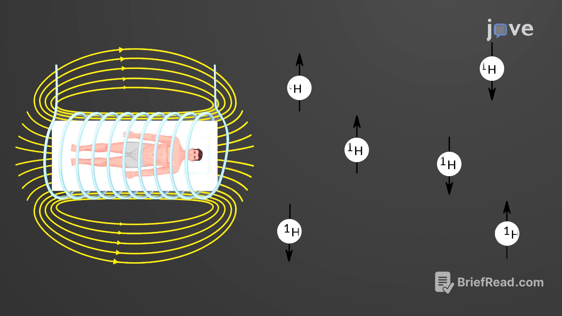
Magnetic Resonance Imaging (MRI) is a noninvasive medical imaging technique that uses magnetic fields and radio waves to produce detailed images of the body. Discovered in the 1930s, the phenomenon involves matter emitting radio signals when exposed to magnetic fields and radio waves. In 1970, Raymond Damadian observed that malignant tissue emitted different signals compared to normal tissue and patented the first MRI scanning device, which was in clinical use by the early 1980s. Early MRI scanners were basic, but advancements in digital computing and electronics improved their precision, especially for tumor detection.
Advantages and Disadvantages of MRI
MRI offers advantages over techniques like computed tomography and X-ray by not exposing patients to radiation. However, MRI scans are more expensive and can cause patient discomfort. The procedure involves powerful electromagnets, requiring shielded scan rooms, and patients must be enclosed in a metal tube for up to thirty minutes, which can be uncomfortable. The machine's noise can also induce anxiety, though "open" MRI scanning helps alleviate these issues. Patients with metallic implants containing iron are not suitable for MRI scans due to the risk of implant dislodgement.
Types of MRI Images and Contrast Media
Different MRI images are produced based on the timing between magnetic pulses and signal interpretation. T1-weighted images show fatty tissues as brighter while suppressing water signals, making them appear darker, whereas T2-weighted images enhance water signals. Contrast media like gadolinium, injected intravenously, improve image quality. Gadolinium, a paramagnetic substance, shortens T1 values in tissues where it accumulates, causing these tissues to appear brighter in T1-weighted images.
Functional and 4D Flow MRI
Functional MRIs (fMRIs) are increasingly used to study brain activity by detecting blood flow concentration in specific areas during various activities, helping scientists understand brain functions, abnormalities, and diseases. More recently, 4D flow MRI provides 3D blood flow images and hemodynamics over time, enabling cardiovascular disease assessment through 3D anatomical and velocity coverage.


![Roller Coaster the Series EP 6 [1/6] The sad farewell of Loft and Pure 😭 รัก ขบวนนี้หัวใจเกือบวาย](https://wm-img.halpindev.com/p-briefread_c-10_b-10/urlb/aHR0cDovL2ltZy55b3V0dWJlLmNvbS92aS9HSUwyeGRjcFJHUS9ocWRlZmF1bHQuanBn.jpg)