TLDR;
This video provides a comprehensive tutorial on the standard radiographic projections of the lumbar spine. It covers the anatomy of the lumbar spine, including the vertebral bodies, intervertebral discs, pedicles, transverse processes, posterior arch, and sacrum. The video also discusses the importance of counting the non-rib bearing segments to ensure accurate identification of the lumbar vertebrae. It highlights the significance of the pars interarticularis and its visualization on oblique projections. Finally, the video explains the orientation of the pedicles in the lumbar spine and how this affects the visualization of the intervertebral foramina on lateral projections.
- The video covers the standard radiographic projections of the lumbar spine, including AP lumbar, AP lumbo pelvic, lateral lumbar, and oblique projections.
- It explains the anatomy of the lumbar spine, including the vertebral bodies, intervertebral discs, pedicles, transverse processes, posterior arch, and sacrum.
- The video emphasizes the importance of the pars interarticularis and its visualization on oblique projections.
- It also discusses the orientation of the pedicles in the lumbar spine and how this affects the visualization of the intervertebral foramina on lateral projections.
Lumbar Spine Anatomy [0:00]
This chapter introduces the standard radiographic projections of the lumbar spine, including AP lumbar, AP lumbo pelvic, lateral lumbar, and oblique projections. It also mentions additional projections like AP angulated spot, PA angulated spot, and lateral lumbo sacral spot projections, which are used to visualize specific areas like the lumbo sacral junction. The chapter emphasizes the importance of understanding the anatomy of the lumbar spine for accurate interpretation of radiographic images.
Basic Radiographic Anatomy [0:54]
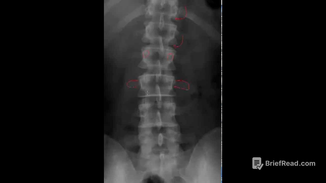
This chapter focuses on the basic radiographic anatomy of the thoraco lumbar region. It highlights the importance of counting the non-rib bearing segments to ensure accurate identification of the lumbar vertebrae. The chapter explains that the presence of five non-rib bearing segments indicates the absence of transitional area anomalies. It also discusses the appearance of the vertebral bodies, intervertebral disc spaces, pedicles, transverse processes, and posterior arch on AP projections. The chapter emphasizes the importance of understanding the anatomy of the lumbar spine for accurate interpretation of radiographic images.
Posterior Arch Anatomy [2:53]
This chapter delves into the anatomy of the posterior arch in the lumbar spine. It explains the different structures within the posterior arch, including the superior and inferior articular processes, pars interarticularis, lamina, and spinous process. The chapter highlights the importance of the pars interarticularis, which is a common site for stress fractures, and explains how it is best visualized on oblique projections. It also discusses the visualization of the facet joints on AP projections.
Sacrum and Other Structures [4:08]
This chapter focuses on the anatomy of the sacrum and other structures visible on AP projections of the lumbar spine. It explains the appearance of the sacral base, sacral iliac joint, anterior and posterior sacral foramina, and the shadow of the soas muscle. The chapter also discusses the potential for spinal infections and other abnormalities to obscure the visualization of the soas shadow. It mentions the possibility of seeing ribs and kidney shadows on AP projections.
Lateral Projection Anatomy [6:24]
This chapter focuses on the anatomy of the lumbar spine as seen on lateral projections. It explains the visualization of the vertebral bodies, pedicles, articular pillars, pars interarticularis, spinous process, and intervertebral disc spaces. The chapter highlights the difficulty in visualizing unilateral pars defects on lateral projections due to the overlapping of structures. It also discusses the visualization of the sacral base, anterior margin of the sacrum, S1-S2 rudimentary disc area, and ribs on lateral projections.
Intervertebral Foramina [9:04]
This chapter focuses on the visualization of the intervertebral foramina on lateral projections of the lumbar spine. It explains the orientation of the pedicles in the lumbar spine and how this allows for direct visualization of the intervertebral foramina on lateral projections. The chapter contrasts this with the cervical spine, where the pedicles are oriented at a 45-degree angle, requiring oblique projections to visualize the intervertebral foramina. It emphasizes the importance of understanding the orientation of the pedicles for accurate interpretation of radiographic images.