TLDR;
This lecture provides an overview of imaging techniques for diagnosing sports injuries, emphasizing their importance in treatment planning, rehabilitation monitoring, and injury prevention. It covers various modalities like X-ray, CT scan, MRI, ultrasound, bone scan, and fluoroscopy, detailing their uses, advantages, and limitations. The lecture also addresses imaging for specific injuries such as fractures, ligament, muscle, tendon, and cartilage damage, as well as concussions and common joint injuries. It concludes by discussing the role of imaging in return-to-play decisions and future trends in imaging technology.
- Imaging is crucial for diagnosing sports injuries, treatment planning, and monitoring rehabilitation.
- Different imaging modalities like X-ray, CT scan, MRI, and ultrasound are used based on the type and severity of the injury.
- Future trends include 3D imaging, AI in interpretation, and portable MRI/ultrasound devices.
Introduction [0:15]
The lecture introduces the importance of imaging techniques in diagnosing sports injuries. Imaging is essential for treatment planning, monitoring rehabilitation progress, and preventing injuries by detecting early signs like stress reactions. It also helps differentiate between acute and chronic conditions.
Overview of Imaging Modalities [1:56]
This section provides a brief overview of various imaging modalities. X-rays are used as a first-line imaging for fractures and joint abnormalities. Musculoskeletal ultrasound is used for dynamic imaging of soft tissue injuries. MRI is best for soft tissue and ligamentous injuries. CT scans are high-resolution imaging for complex fractures. Bone scans are used to detect stress fractures and metabolic bone diseases. Fluoroscopy is used for real-time imaging for guided procedures.
X-Rays [3:59]
X-rays are ideal for detecting fractures, dislocations, and joint abnormalities. They are quick, cost-effective, and widely available, even in portable forms. However, they have limitations in visualizing soft tissues. X-rays are commonly used in high-impact sports like football, basketball, and rugby. Standard views include anterior-posterior, lateral, and oblique, with stress radiographs used for assessing ligamentous stability. Weight-bearing views can detect joint instability and cartilage loss.
CT Scan [5:10]
CT scans provide cross-sectional imaging with high detail, useful for complex fractures of the face, pelvis, and spine. They can detect bony lesions and small fractures not visible on X-rays but involve higher radiation exposure.
MRI [5:43]
MRI is considered the gold standard for soft tissue injuries, excellent for detecting ligament tears (ACL, PCL, MCL, LCL), meniscal injuries, muscle strains, and cartilage damage. It has no radiation exposure but is expensive and time-consuming. Specific sequences like T1-weighted (anatomical details), T2-weighted (fluid and edema), STIR (bone marrow edema, stress fractures), and DWI (acute injuries) are used.
Ultrasound Imaging [6:48]
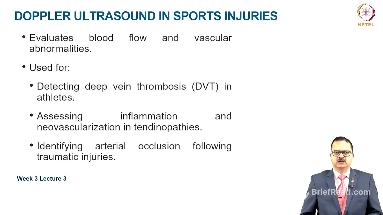
MSK ultrasound is portable, cost-effective, and offers real-time imaging, best for tendon, ligament, and muscle injuries. It allows dynamic assessment of movement abnormalities and is used for guiding injections like corticosteroids and PRP. Doppler ultrasound evaluates blood flow, detecting vascular abnormalities, deep vein thrombosis, inflammation, neovascularization in tendinopathies, and arterial occlusion following traumatic injuries.
Bone Scan [7:56]
Bone scans are highly sensitive for detecting stress fractures, bone infections, and metabolic disorders affecting the bone. They are useful in endurance athletes for early-stage stress fracture detection, but lack specificity, making MRI the preferred choice in most cases.
Fluoroscopy [8:27]
Fluoroscopy provides real-time X-ray imaging for dynamic joint assessments and interventional procedures, such as guided joint injections. It can also be used for stress testing of ligaments and identifying joint instability, but MSK ultrasound is now more commonly used for these procedures.
Imaging for Specific Injuries [9:08]
X-rays are the first-line modality for fractures, with CT scans useful for complex fractures. MRI can detect stress fractures if X-rays are inconclusive. For ligament injuries, MRI is best, while ultrasound assesses superficial injuries dynamically, and stress X-rays evaluate ligament instability. In muscle and tendon injuries, ultrasound is best for acute muscle tears and tendinopathy, but MRI is the gold standard for deep muscle injuries and complex tendon tears. Doppler ultrasound evaluates vascularity in chronic tendinopathies. MRI with specialized sequences is used for cartilage injuries, CT arthrography detects small cartilage defects, and X-rays assess osteoarthritis progression. MRI is most sensitive for early stress fracture detection, with bone scans as an alternative if MRI is unavailable.
Imaging for Specific Conditions [11:12]
CT scans are the first-line modality for acute intracranial injuries in concussions and head trauma. MRI detects post-concussion syndrome and chronic symptoms, with diffusion tensor imaging emerging for subtle brain injuries. For shoulder injuries, X-rays assess fractures and dislocations, MRI detects rotator cuff tears and labral injuries, and MSK ultrasound assesses impingement syndromes dynamically. In knee injuries, MRI is best for meniscus, ligament, and cartilage injuries, while X-rays have limited use except for fractures and arthritis, and ultrasound evaluates bursitis and effusions. X-rays are excellent for first-line imaging of ankle and foot injuries. MRI identifies ligament injuries and stress fractures, and ultrasound assesses plantar fasciitis and ligament injuries. For hip and groin injuries, X-rays detect fractures and impingement syndromes, MRI is best for labral tears and muscle injuries, and ultrasound evaluates sports hernias and adductor injuries.
Role of Imaging in Return-to-Play (RTP) Decisions [13:12]
Imaging helps monitor healing progression with serial imaging, provides functional imaging for dynamic assessments, and offers objective evaluation to prevent premature return and further reinjury.
Future Trends in Imaging Technology [13:40]
Advances include three-dimensional imaging reconstruction for better visualization, artificial intelligence in imaging interpretation, and portable MRI or on-field ultrasound devices for on-field assessment.
Limitations and Considerations for Imaging [14:11]
Cost and accessibility are limitations for advanced imaging modalities. Radiation exposure is a concern for young athletes. Over-reliance on imaging versus clinical examination and correlation should be avoided; imaging should confirm a diagnosis rather than be the sole basis for it.
Conclusion [14:43]
Imaging is crucial for accurate sports injury diagnosis and management. The choice of modality depends on injury type and severity, and future advancements will combine diagnostic precision to enhance athlete care.