TLDR;
This lecture series focuses on the development of the central nervous system and the stages of fertilization and embryonic development. Key points include the fertilization process, stages of cell division, and the differentiation of the inner and outer cell masses.
- Understanding fertilization and the stages of early development.
- The significance of hormonal influences in implantation and embryo development.
Introduction to Neuroanatomy [0:00]
The lecture begins with an overview of neuroanatomy and its significance in medicine. Neuroanatomy can be daunting for some medical professionals, but a step-by-step approach makes it easier to understand. The focus shifts toward the development of the central nervous system, starting from the zygote through to embryo and fetus stages.
Fertilization Process [0:36]
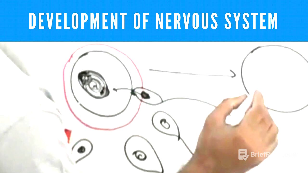
The process begins with the release of an ovum from the Graafian follicle into the fallopian tube, where it may encounter sperm. When the sperm penetrates the ovum, it triggers the completion of the second meiotic division in the ovum, resulting in a definitive ovum with one polar body. This ovum now contains genetic material from both parents, leading to the formation of two pronuclei: female from the ovum and male from the sperm.
Zygote to Blastocyst Development [6:44]
Following fertilization, the zygote undergoes mitotic division, first forming a two-cell stage and then proceeding to a four-cell stage, eventually becoming a blastula. Each division decreases the overall cell size while the zona pellucida remains intact. As the blastula develops, it transitions into a morula, defined by a mass of cells that will later differentiate into an inner cell mass and outer cell mass.
Blastocyst Structure and Functions [12:43]
The blastocyst forms when fluid accumulates within the outer cell mass, leading to a fluid-filled cavity. The inner cell mass is now referred to as the embryoblast, while the outer mass is known as the trophoblast. The trophoblast develops into a layer that will ultimately become the placenta, providing nourishment to the developing embryo.
Implantation Process [19:34]
As the blastocyst reaches the uterine cavity, the villi—which have formed from the trophoblast—attach to the endometrium. The trophoblast releases enzymes that facilitate implantation, which is crucial for establishing the pregnancy. The hormonal environment of the endometrium, influenced by progesterone, becomes highly conducive for implantation.
HCG and Maintenance of Pregnancy [26:14]
After implantation, the trophoblast produces human chorionic gonadotropin (HCG), which signals the corpus luteum to continue producing progesterone, thus maintaining the endometrium. This hormonal support ensures that menstrual cycles do not resume, allowing for the growth and development of the embryo within the uterine environment.
Early Embryonic Development [28:34]
In the second week following implantation, the inner cell mass begins to differentiate into two layers. By this time, the embryoblast has attached itself firmly to the endometrium, supported by high levels of progesterone which maintain the pregnancy and prepare the uterine environment for the developing embryo.