TLDR;
This video provides an overview of the four main types of animal tissues: epithelial, nervous, connective, and muscle. It explains the structure, function, and location of various subtypes within each tissue category, offering tips on how to identify them under a microscope.
- Epithelial tissue serves as a protective lining.
- Nervous tissue transmits electrochemical signals.
- Connective tissue supports and stabilizes body structures.
- Muscle tissue enables movement.
Introduction to Animal Tissues [0:01]
The video introduces the four main types of animal tissues: connective, epithelial, muscle, and nervous tissue. Each tissue type has specific functions linked to its shape. The video will cover the specifics of each tissue, including their appearance and how to identify them, using micrograph pictures and examples of how they might be presented in tests or exams.
Epithelial Tissue: Structure and Types [0:38]
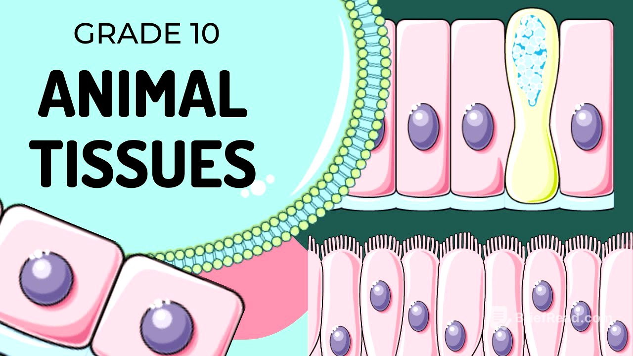
Epithelial tissue, a simple tissue, is often one layer thick but can also be multi-layered (stratified). It serves as a protective layer held together by a basement membrane, a thin membrane that acts as a sticky surface for cells to attach. Epithelial tissue has very few intercellular spaces because its function is to protect and line internal and external surfaces of the body. A thin layer of connective tissue sits under the basement membrane to connect the epithelial tissue to underlying tissues like fat or muscle. Key features include geometric shapes and nuclei that can be circular or elongated. The three basic shapes of epithelial tissue are squamous, columnar, and cuboidal.
Squamous, Columnar, and Cuboidal Epithelium [3:33]
Squamous epithelium consists of thin, irregularly shaped, and tightly compacted cells with flattened nuclei. It is found in the lining of the mouth, alveoli, and the surface of the skin, and it renews quickly. Columnar epithelium is composed of elongated, rectangular cells with oval-shaped nuclei, often positioned upwards. These cells can be ciliated, increasing surface area for absorption and secretion of mucus, and are found in the intestines and gall bladder. Cuboidal cells are square-shaped with very spherical nuclei, typically lining tubes and functioning in absorption and secretion, such as in sweat glands, the thyroid gland, and kidney tubules.
Nervous Tissue and Neuron Types [7:58]
Nervous tissue conducts electrochemical signals between organs and the brain. There are three types of nervous tissue cells: sensory neurons, motor neurons, and relay neurons (interneurons). All nerve cells have dendrites, a cell body, an axon, and axon terminals. Sensory neurons sense information, with the cell body sitting off to the side. Motor neurons are responsible for movement, with the cell body in the center of the dendrites. Relay neurons (interneurons) relay information between sensory and motor neurons, found mostly in the spinal cord and brain, with the cell body in the center of the nerve cell. The location of the cell body determines the location of the axon versus the dendrites.
Connective Tissues: Types and Functions [13:58]
Connective tissues support, stabilize, and protect the body's organs. They consist of cells surrounded by a fluid or matrix, which can be liquid, semi-liquid, or solid. The six major types of connective tissues are areolar tissue, fibrous connective tissue, cartilage, blood, adipose tissue, and bone tissue.
Areolar, Fibrous, and Cartilage Connective Tissues [14:59]
Areolar tissue binds epithelium and is found under epithelial tissue and between organs, attaching them loosely. Its matrix is made of collagen and elastic fibers. Fibrous connective tissue is a dense network of non-elastic collagen, found in tendons and ligaments, providing strength and flexibility. Cartilage prevents friction and consists of chondrocytes in a semi-fluid matrix, providing a smooth, glossy appearance and absorbing shock, found between bones and at joints.
Blood, Adipose, and Bone Connective Tissues [18:35]
Blood is the only liquid connective tissue, consisting of lymphocytes (white blood cells for immunity), erythrocytes (red blood cells for carrying gases), and platelets (for blood clotting). It transports waste, nutrients, and hormones within the circulatory system. Adipose tissue is made of fat cells, storing excess energy and providing insulation, found around organs and under the skin. Bone tissue provides the framework for the body, made up of osteocytes that secrete a solid matrix, arranged in Haversian systems within the skeletal system.
Muscle Tissue: Skeletal, Smooth, and Cardiac [21:53]
Muscle tissue falls into three categories: skeletal, smooth, and cardiac muscle. Skeletal muscle has striations and multiple nuclei, involved in voluntary movement and attached to bones. Smooth muscle is non-striated, responsible for involuntary movement, and found in the digestive system and blood vessels. Cardiac muscle, found only in the heart, is a network of branched muscle fibers with faint striations, responsible for involuntary movement and capable of moving on its own without brain instructions.
Summary of Tissue Types and Functions [26:11]
The video summarizes the four types of tissues: epithelial, nervous, connective, and muscle. Epithelial tissue lines surfaces and provides protection. Nervous tissue transports electrochemical signals. Connective tissue holds tissues together. Muscle tissue allows movement. Specific types of epithelium (squamous, columnar, cuboidal), neurons (sensory, motor, interneurons), and connective tissues (areolar, fibrous, blood, bone, adipose) are reviewed, along with the characteristics and functions of skeletal, smooth, and cardiac muscle.