TLDR;
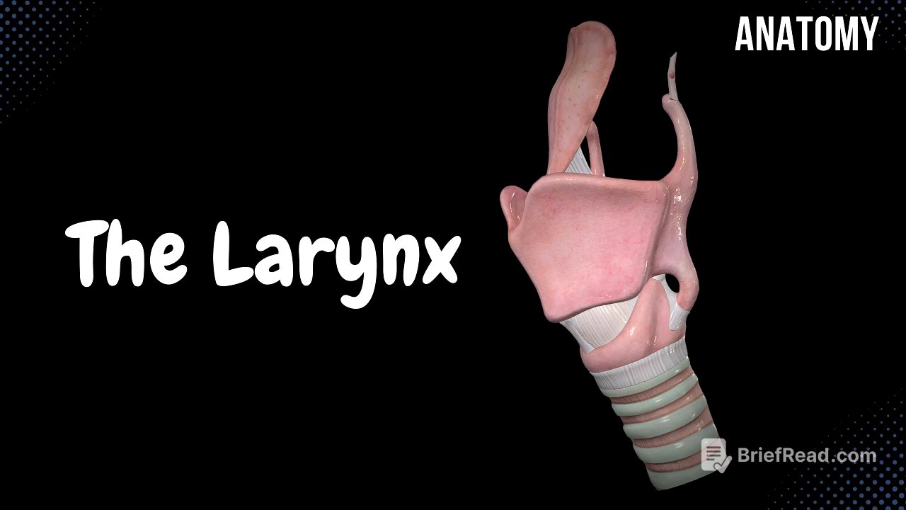
Alright, so this video is all about the anatomy of the larynx, or the voice box. It covers the location and function of the larynx, the cartilages that make it up (thyroid, cricoid, epiglottis, arytenoid, corniculate, and cuneiform), the ligaments and joints that hold it all together, the layers of the laryngeal wall (tunica mucosa, tela submucosa, cartilage and muscle layer, and tunica adventitia), and the different parts of the laryngeal cavity. Key takeaways include understanding how the vocal cords work for phonation and the importance of the rima glottidis.
- Larynx location and function
- Cartilages, ligaments, and joints of the larynx
- Layers of the laryngeal wall
- Laryngeal cavity and phonation
Introduction [0:00]
The video introduces the anatomy of the larynx as part of the respiratory system, which includes the nose, pharynx, larynx, trachea, bronchi, and lungs. The video will cover the orientation of the larynx, its cartilages, ligaments, joints, walls, and the laryngeal cavity.
Orientation of the Larynx [0:50]
The larynx is located between the hyoid bone and the trachea, in front of the esophagus, spanning from the 4th-5th to the 6th-7th cervical vertebrae. Its functions include serving as an air passage and producing sound through phonation, where vocal cords rub together to create different pitches. The video shows anterior and posterior views of the larynx, highlighting its position relative to the hyoid bone and trachea.
Cartilage of the Larynx [1:47]
The larynx is made up of six cartilages: three unpaired (epiglottis, thyroid, and cricoid) and three paired (arytenoid, corniculate, and cuneiform). The unpaired cartilages include the epiglottis, thyroid cartilage, and cricoid cartilage. The paired cartilages are the arytenoid, corniculate, and cuneiform cartilages.
Thyroid Cartilage [2:42]
The thyroid cartilage consists of two plates or laminas (right and left) that meet in the middle to form the laryngeal prominence, also known as the Adam's apple. It has two processes: the superior horn, which points towards the hyoid bone, and the inferior horn, which points towards the cricoid cartilage.
Cricoid Cartilage [3:30]
The cricoid cartilage, located below the thyroid cartilage, has an arch anteriorly and a plate posteriorly with two important surfaces. These surfaces include the arytenoid articular surface, where the arytenoid cartilage connects to form a joint, and the thyroid articular surface, which forms a joint with the inferior horn of the thyroid cartilage.
Epiglottis [4:06]
The epiglottis is located behind the thyroid cartilage and hyoid bone, attaching to the thyroid cartilage via ligaments. Its primary function is to close off the respiratory system during swallowing and open it during breathing.
Arytenoid Cartilages [4:31]
The arytenoid cartilages are paired and located above the cricoid cartilage. They have a triangular shape with an apex on top and a base with two processes. The anterior process is the vocal process, attaching to the vocal ligament, and the posterior process is the muscular process, serving as an attachment point for muscles. The base is called the cricoid articular surface.
Corniculate Cartilage [5:11]
The corniculate cartilages are located on top of the arytenoid cartilages and primarily serve as attachment points for certain muscles.
Cuneiform Cartilages [5:27]
The cuneiform cartilages are small, elongated pieces of elastic cartilage located in the aryepiglottic fold, which lines the entrance of the larynx. They form a tubercle called the cuneiform tubercle, visible during examination of the larynx.
Ligaments and Joints [6:00]
The connections in the larynx are divided into continuous and discontinuous articulations. Continuous connections include cartilaginous (synchondroses) and fibrous connections (membranes or ligaments), while discontinuous connections are synovial joints. The larynx has one cartilaginous connection between the corniculate and arytenoid cartilages. Fibrous connections include the thyrohyoid membrane (median and lateral), cricothyroid membrane (median cricothyroid ligament and lateral cricothyroid ligament or conus elasticus), cricotracheal ligament, thyroepiglottic ligament, and hyoepiglottic ligament. The larynx has two synovial joints: the cricothyroid articulation (between the inferior horn of the thyroid cartilage and the cricoid cartilage) and the cricoarytenoid articulation (between the base of the arytenoid cartilage and the cricoid cartilage).
Laryngeal Wall [9:32]
The laryngeal wall consists of four layers: the tunica mucosa, tela submucosa, a layer of cartilages and muscles, and the tunica adventitia.
Tunica Mucosa [10:19]
The tunica mucosa is the innermost lining of the larynx. The vestibular folds are lined by respiratory epithelium with cilia for filtering air, while the vocal folds are lined by stratified squamous epithelium to withstand strain. The tunica mucosa also contains laryngeal glands for lubrication and small lymph nodules for immunity.
Tela Submucosa [11:31]
In some areas, the fibrous membranes form a fibroelastic membrane, which plays a key role in speech. There are two fibroelastic membranes: the quadrangular membrane (between the vestibular folds and the epiglottis, with the upper margin forming the aryepiglottic fold and the lower margin forming the vestibular ligament) and the conus elasticus or lateral cricothyroid ligament (connected to the cricoid, arytenoid, and thyroid cartilages, with the upper margin forming the vocal ligament).
Muscles of the Larynx [13:06]
The muscles of the larynx are grouped according to their function: muscles that open and narrow the laryngeal inlet, muscles that open and narrow the rima glottidis (the space between the vocal folds), and muscles that act on the vocal cord itself. The vocal ligament is located within the mucus membranes, and together they form the vocal cord. Muscles like the cricothyroid tense the vocal cord, while muscles like the vocalis decrease the tension.
Tunica Adventitia [15:46]
The tunica adventitia is a tough connective tissue covering around the larynx, consisting mainly of dense collagen fibers.
Laryngeal Cavity [16:03]
The laryngeal cavity extends from the laryngeal inlet to the lower border of the cricoid cartilage and is divided into three parts: the laryngeal vestibule (from the laryngeal entrance to the vestibular folds), the glottis (between the vestibular folds and the vocal cords), and the infraglottic cavity (from the vocal cords to the lower border of the cricoid cartilage). The glottis is divided into an anterior 3/5 intermembranous part and a posterior 2/5 intercartilaginous part. The laryngeal ventricles act as resonators during speech.


