TLDR;
This video provides an overview of the integumentary system, which includes the skin, hair, nails, and glands. It details the structure and function of the skin, discussing the epidermis and dermis layers, as well as the different cell types and layers within the epidermis. The video also covers hair follicles, nail structure, and the various types of glands found in the skin, such as sweat glands and sebaceous glands, and their functions in protection, temperature regulation, and sensory response.
- The skin is composed of two main layers: the epidermis and the dermis.
- Hair follicles produce hair, which consists of the medulla, cortex, and cuticle.
- Nails are made of hard keratin and include the free edge, body, and proximal root.
- Glands in the integumentary system include sweat glands (eccrine and apocrine) and sebaceous glands.
Introduction to the Integumentary System [0:00]
The integumentary system, the body's outermost system, comprises the skin and its derivatives, such as glands, hair, and nails. The skin acts as a protective barrier against external elements, preventing bacteria from entering and retaining water and heat. The video will explore the structure and function of the skin and its components.
Skin Structure: Epidermis and Dermis [0:42]
The skin consists of two primary regions: the epidermis, a thin outer layer, and the dermis, a thicker inner layer. The dermis is a vascularised layer of fibrous connective tissue, while the epidermis, made of epithelial tissue, is avascular and receives nutrients through diffusion from the dermis. Below the dermis is the subcutaneous layer, or hypodermis, which is mainly adipose tissue that anchors the skin to underlying structures.
Epidermis: Layers and Cell Types [1:38]
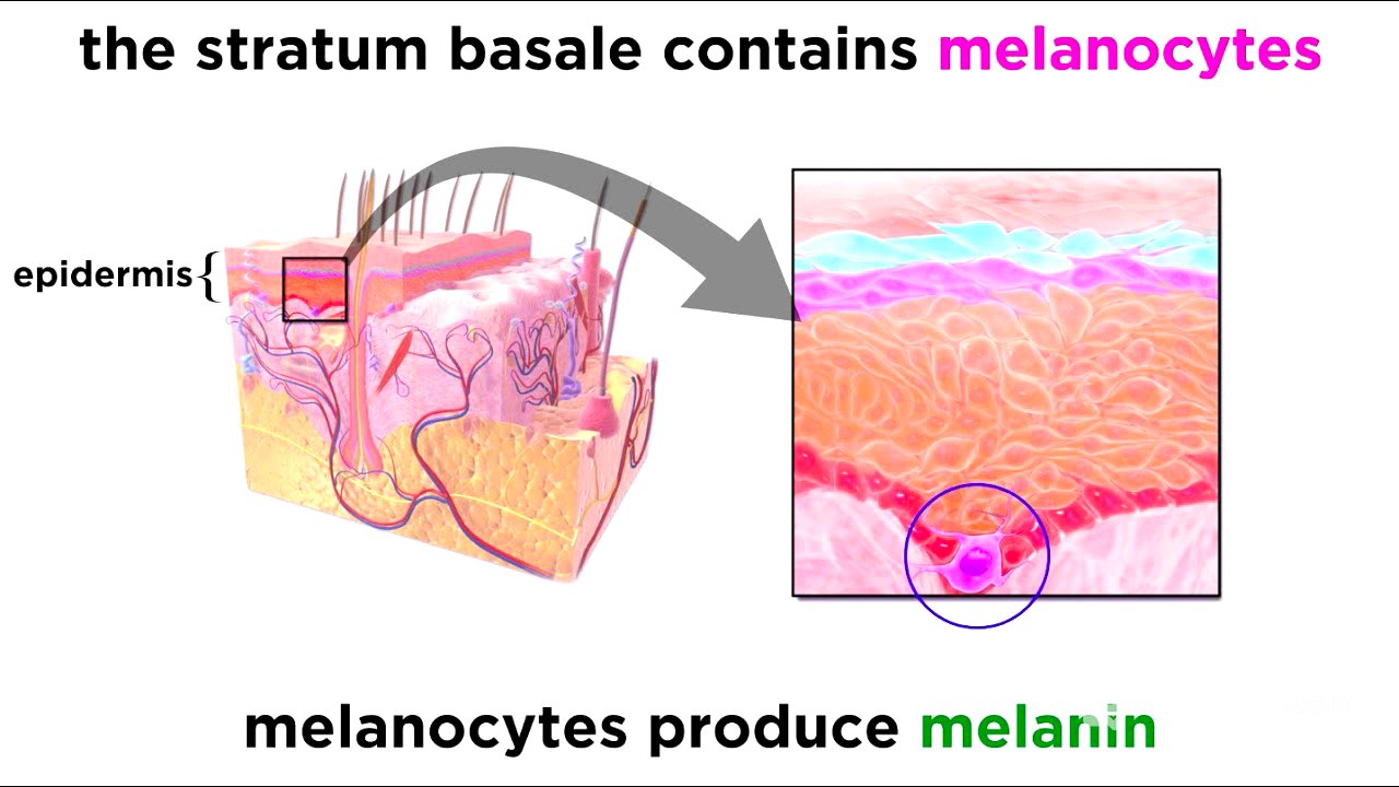
The epidermis is composed of keratinised stratified squamous epithelium, consisting of four cell types in five layers. The deepest layer, the stratum basale, contains rapidly dividing keratinocytes that regenerate dead skin and produce keratin. This layer also includes melanocytes, which produce melanin, and tactile cells, which act as sensory receptors for touch. Above this is the stratum spinosum, containing cells with a weblike system of intermediate filaments and dendritic cells that activate the immune system. The stratum granulosum is where keratinisation begins, as cells fill with keratin and die. The stratum lucidum is a clear layer of dead keratinocytes, and the outermost stratum corneum consists of many layers of dead, anucleated cells filled with keratin, protecting the living cells beneath.
Dermis: Papillary and Reticular Layers [4:55]
The dermis is made of strong, flexible connective tissue containing nerves, blood vessels, and hair follicles. It has two sections: the papillary layer and the reticular layer. The papillary layer, made of areolar connective tissue, features dermal papillae that project into the epidermis and contain tactile cells. In areas of high friction, these papillae sit on dermal ridges, enhancing gripping ability and forming unique fingerprints. The reticular layer, comprising most of the dermis, is made of dense fibrous connective tissue.
Skin Pigmentation [6:34]
Skin colour is determined by melanin, produced by melanocytes in the stratum basale, which protects the skin from ultraviolet radiation. Other pigments contributing to skin colour include carotene (yellow-orange) and haemoglobin (red when oxygenated).
Skin Appendages: Hair Structure and Follicles [7:11]
Hair, a skin appendage, is a flexible strand made of dead, keratinised cells produced by hair follicles. A hair consists of a root (where keratinisation occurs) and a shaft (where keratinisation is complete). The hair itself has three layers: the medulla (containing large cells and soft keratin), the cortex (flattened cells), and the cuticle (overlapping, keratinised cells). The hair follicle is a pocket extending from the epidermis into the dermis, expanding at the deep end to form a hair bulb. Nerve endings attach to the bulb, acting as receptors, and a hair papilla supplies nutrients for hair growth.
Hair Follicle Structure and Function [9:24]
The hair follicle wall consists of the peripheral connective tissue sheath (from the dermis), the glassy membrane (from the basal lamina), and the epithelial root sheath (from the epidermis). Cells in the hair matrix divide, pushing existing cells upwards and causing hair to grow. Each follicle has an arrector pili, a muscle bundle that contracts to cause goose bumps. Hair can be vellus (pale and fine) or terminal (darker and coarse).
Nails: Structure and Growth [10:45]
Nails, modifications of the epidermis, contain hard keratin. Each nail has a free edge, a body, and a proximal root embedded in the skin. The nail sits on the nail bed and grows from the nail matrix. Skin folds called nail folds sit on the borders of the nail, with the eponychium extending onto the nail. The hyponychium is at the edge of the finger.
Glands: Sweat, Ceruminous, Mammary, and Sebaceous [11:53]
The integumentary system includes various glands. Sweat glands (sudoriferous glands) are of two types: eccrine (merocrine) sweat glands, which secrete sweat through pores, and apocrine sweat glands, which secrete fat and protein components, causing body odour. Ceruminous glands produce earwax, and mammary glands produce breast milk. Sebaceous glands (oil glands) secrete sebum, which lubricates hair and skin, slows water loss, and kills bacteria.
Conclusion: Functions of the Integumentary System [13:31]
The integumentary system, consisting of skin, hair, nails, and glands, acts as a barrier, separating the inside of the body from the outside. It repairs quickly, regulates body temperature, and responds to external stimuli.