TLDR;
Ultrasonography is a non-invasive imaging technique using high-frequency sound waves to visualize internal body structures without ionizing radiation. It's used across medical fields to study heart function, blood flow, and fetal development. The procedure involves a transducer emitting sound waves that bounce back to create real-time images. Key applications include diagnostics, obstetrics/gynecology, vascular studies, and musculoskeletal imaging.
- Non-invasive and safe imaging technique.
- Used for diagnostics, obstetrics, vascular, and musculoskeletal studies.
- Image quality depends on the operator and is limited by bone, gas, and excess subcutaneous fat.
Ultrasonography: Overview and Procedure
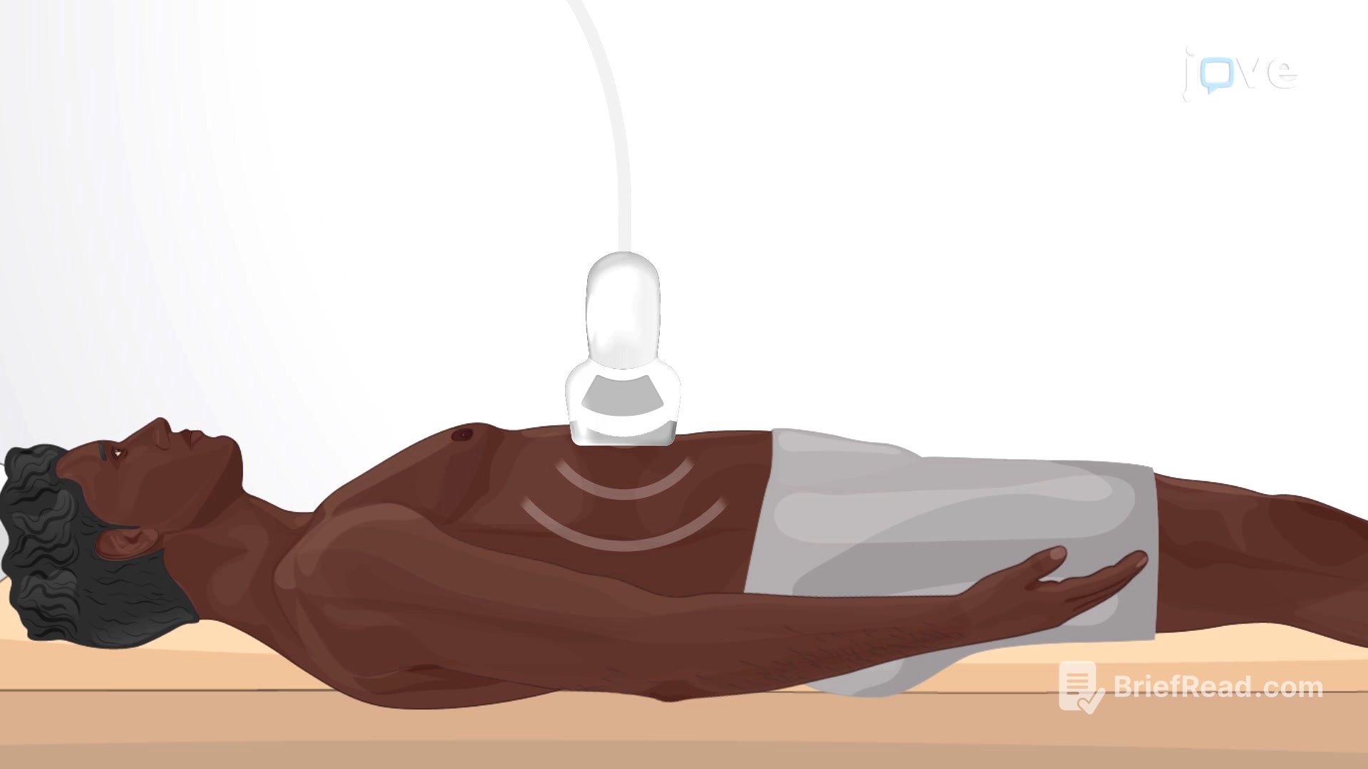
Ultrasonography is an imaging method that uses high-frequency sound waves to visualize internal body structures. It is a non-invasive and safe procedure because it does not use ionizing radiation. This makes it suitable for various medical applications, including studying heart function, blood flow in the neck or extremities, gallbladder disease, and fetal growth. During the procedure, a transducer emits sound waves that bounce back from tissues and structures, creating real-time images on a computer monitor.
Applications of Ultrasonography
Ultrasonography is used for diagnostic purposes to identify abnormalities like tumors, cysts, gallstones, and fluid collections, and to evaluate organs such as the liver, kidneys, bladder, uterus, and prostate. In obstetrics and gynecology, it monitors pregnancy, assesses fetal development, determines gestational age and position, detects multiple pregnancies, and evaluates the uterus and placenta. It also examines blood vessels to detect blockages or abnormalities, using Doppler ultrasound to evaluate blood flow direction and velocity. Additionally, ultrasonography evaluates the musculoskeletal system, identifying injuries, inflammation, and guiding needle placements for interventions.
Limitations of Ultrasonography
The main disadvantage of ultrasonography is that the image quality is heavily operator-dependent. Ultrasonography cannot penetrate bone and gas, which limits its use in certain areas of the body. Additionally, excess subcutaneous fat, common in obese individuals, affects the image quality by attenuating the echo, which limits clear visualization.