TLDR;
Positron Emission Tomography (PET) is a medical imaging technique using radiopharmaceuticals to visualize the body's functions. It involves injecting a radiotracer into the patient, which emits positrons that interact with electrons, producing detectable gamma radiation. This radiation is converted into detailed images showing organ function, nutrient metabolism, and blood flow. PET scans are used to diagnose various conditions, including heart disease, cancer spread, brain abnormalities, and thyroid disease. They are often combined with CT or MRI scans for enhanced visualization and interpretation.
- PET scans use radiopharmaceuticals to visualize body functions.
- Radiotracers emit positrons, producing gamma radiation that is detected and converted into images.
- PET is used to diagnose heart disease, cancer, brain abnormalities, and thyroid disease.
- PET scans are often combined with CT or MRI for better visualization.
Positron Emission Tomography
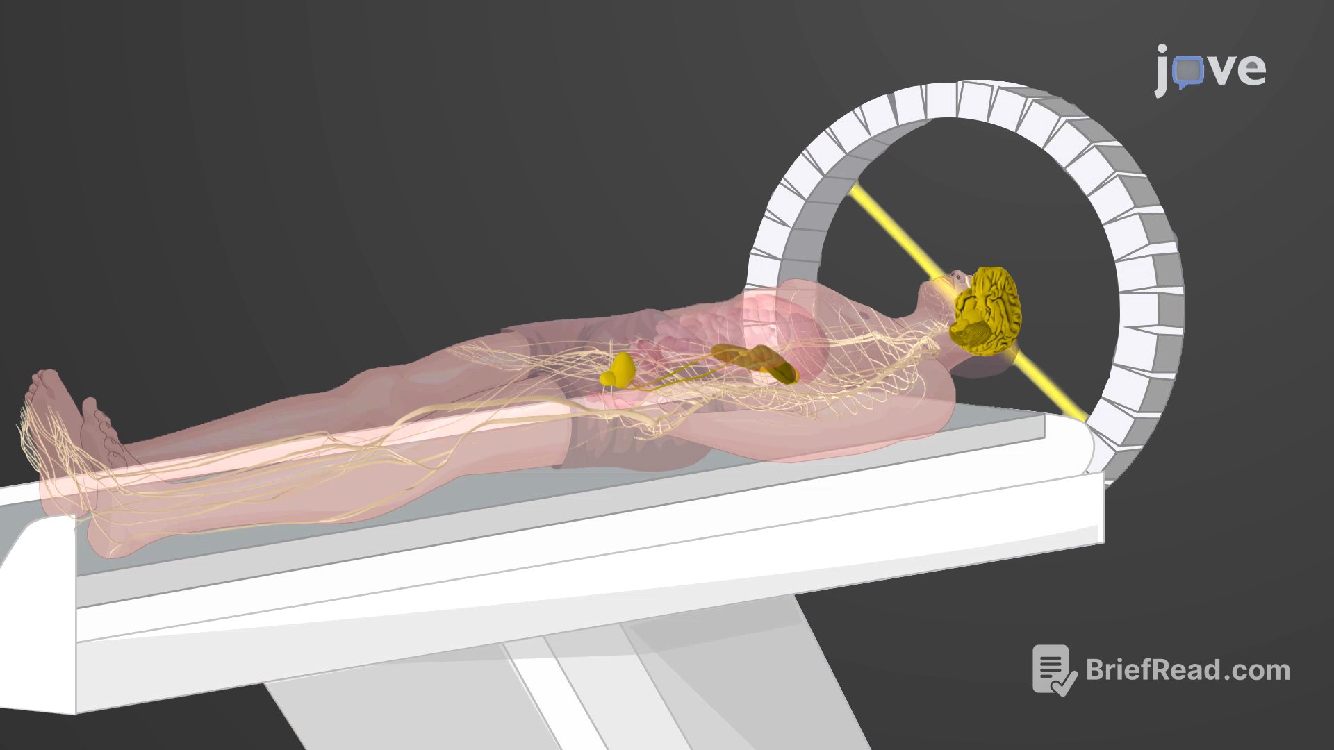
Positron Emission Tomography (PET) is a medical imaging technique that uses radiopharmaceuticals, which are substances emitting short-lived radiation. The first PET scanner was introduced in 1961, but it was the combination with radiopharmaceuticals 15 years later that revolutionized its potential. A key requirement is a positron-emitting radioisotope, produced in a cyclotron and attached to a substance used by the body part being examined. This "tagged" compound, or radiotracer, is administered to the patient, and its use by the tissue reveals the function of the organ or body area. For example, F-18, produced by proton bombardment of 18O, is incorporated into fludeoxyglucose, a glucose analog. The body's use of FDG provides critical diagnostic information, such as revealing cancers due to their different glucose usage compared to normal tissues. The emitted positrons interact with nearby electrons, producing gamma radiation, which is detected by the scanner and converted into a detailed, three-dimensional color image showing the body's function. Different levels of gamma radiation produce varying brightness and colors, which radiologists interpret. Radioactive iodine can monitor the thyroid, and radioactive gallium can be used for cancer imaging. The main advantage of PET is its ability to illustrate the physiologic activity, including nutrient metabolism and blood flow, of the targeted organs, unlike CT and MRI scans that only show static images. PET is widely used to diagnose conditions like heart disease, cancer spread, brain abnormalities, bone disease, and thyroid disease. It can also locate active brain regions during specific activities. PET scans are now commonly performed with CT or MRI scans for improved data visualization and interpretation.