TLDR;
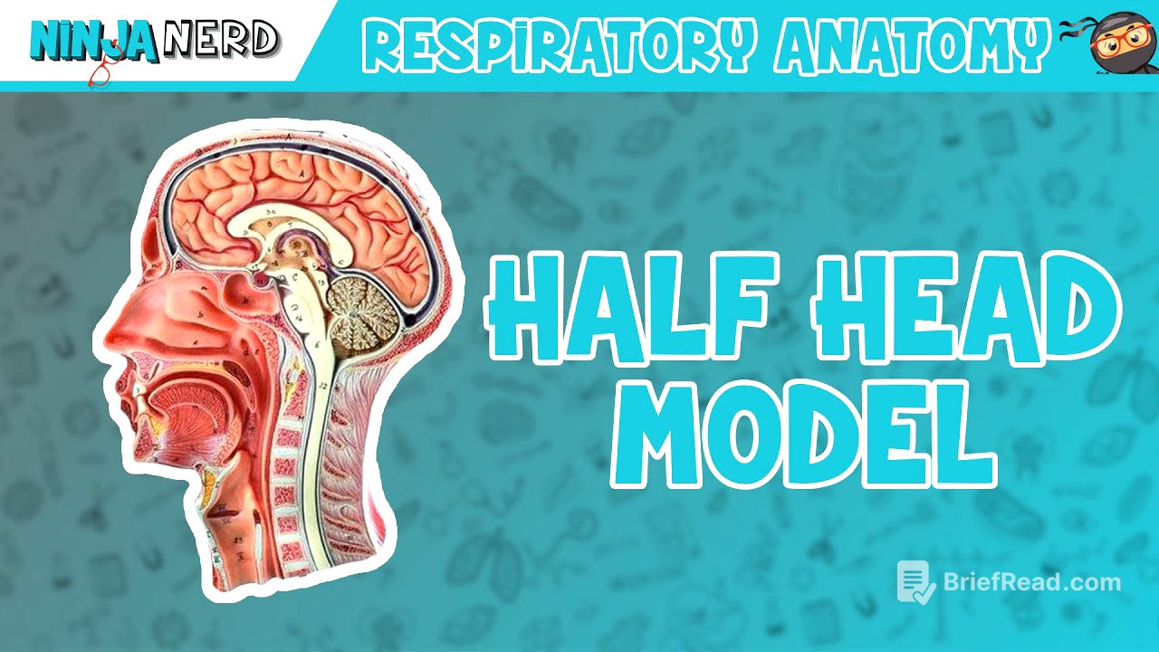
This video provides a detailed overview of the respiratory system's structures using a half-head model. It covers the nasal cavity, pharynx, and larynx, explaining the function and importance of each component. Key points include the roles of the nasal conchae in air humidification and filtration, the three sections of the pharynx (nasopharynx, oropharynx, laryngopharynx) and their tissue composition, and the structure of the larynx with its epiglottis and vocal cords.
- Nasal cavity structures (external nares, vestibule, nasal conchae, meatuses) and their functions in air conditioning and filtration.
- Pharynx divisions (nasopharynx, oropharynx, laryngopharynx) and their respective tissue types and functions.
- Larynx components (epiglottis, vocal cords, thyroid cartilage, cricoid cartilage) and their roles in breathing and phonation.
Introduction to Respiratory System Structures [0:06]
The video begins by introducing the structures of the respiratory system using a half-head model. The initial focus is on the nasal cavity, specifically the vestibule, which is the inner area of the nasal cavity. The external nares serve as the entry point for air, leading into the nasal cavity.
Nasal Cavity Details [0:36]
The nasal cavity is lined with pseudo stratified ciliated columnar epithelial tissue, which is crucial for warming, moistening, filtering, and humidifying incoming air. Cilia in this tissue help in the movement of mucus. Vibrissae, or nose hairs, also play a role in filtering particles entering the nose. The nasal cavity includes the superior, middle, and inferior nasal conchae, with corresponding meatuses (superior, middle, and inferior) in between. These structures turbinate the air, increasing its contact with the mucosal surface to enhance humidification, warming, and filtering.
Posterior Nasal Apertures and Surrounding Structures [2:22]
As air passes through the nasal cavity and its structures, it reaches the posterior nasal apertures, marked by the internal nares. The Ferengi tympanic tube (auditory or eustachian tube) connects to the middle ear, aiding in pressure equalization. Around this tube is lymphatic tissue known as the tubule tonsils. The pharyngeal tonsils are located in the posterior aspect of the nasal cavity.
Paranasal Sinuses and Palate [3:40]
The frontal sinus, part of the frontal bone, is one of the paranasal sinuses. The sphenoid sinus is located near the pituitary gland in the sella turcica. The floor of the nasal cavity, which forms the roof of the oral cavity, consists of the hard palate and the soft palate. The soft palate extends into the uvula. The soft palate, containing muscles like the tensor villi palatini and pelota glossus, elevates during swallowing to block the nasal cavity, preventing food from entering.
Arches and Tonsils [5:26]
The pilato pharyngeal arch and the Pilato Glaus arch are significant structures in this region. The space between these arches is called the fawzi's, which houses the Palatine tonsils. Additionally, the lingual tonsils are located at the back of the tongue.
Pharynx Divisions [5:58]
The pharynx is divided into three sections: the nasopharynx, oropharynx, and laryngopharynx, each with distinct tissue types and functions. The nasopharynx, lined with pseudo stratified ciliated columnar epithelial tissue, primarily handles air. The oropharynx and laryngopharynx are made up of stratified squamous epithelial tissue, suitable for contact with food, fluids, and air, providing resistance against abrasion and friction.
Larynx and Epiglottis [7:46]
The laryngeal pharynx directs air into the larynx. The esophagus, with its upper esophageal sphincter, remains constricted until swallowing occurs. The epiglottis, made of elastic cartilage, is crucial for flexibility and recoil ability. During swallowing, the epiglottis blocks the larynx, directing food into the esophagus. A ligament connects the epiglottis to the hyoid bone, known as the high o epiglottal ligament.
Vocal Cords and Trachea [9:37]
The laryngeal Inlet marks the entry into the larynx. Within the larynx, there are false and true vocal cords. The true vocal cords vibrate as air is pushed out, producing sound. The space between the true vocal cords is the glottis. Past the true vocal cords and the cricoid cartilage lies the trachea.
Cartilage Structures of the Larynx [11:57]
The cricoid cartilage is the only cartilage that completely encircles the larynx. The original cartilage is covered by original muscles, which control the tension of the vocal cords. The hyoid bone is connected to the thyroid cartilage via the tyro hyoid ligament. The thyroid cartilage forms the laryngeal province, or Adam's apple, which is more prominent in males due to testosterone's influence on hyaline cartilage production during puberty. The cricothyroid ligament connects the cricoid and thyroid cartilages.
Arytenoid Cartilage and Conclusion [14:02]
The arytenoid cartilage, along with the core niculae, tanned, and cuneiform cartilages, are better visualized in other models. These structures, along with associated muscles, are important for controlling vocal cord tension. The video concludes by summarizing the covered information and encouraging viewers to like, comment, and subscribe.