TLDR;
This video explains microsporogenesis, the process of pollen formation inside the anther of a flower. It details the structure of the anther, the different layers within it, and the development of microspores from sporogenous cells. The video also covers the importance of meiosis in producing haploid microspores and the role of the tapetum in nourishing developing cells and releasing microspores.
- The stamen, the male reproductive part of a flower, consists of the filament and anther.
- Microsporogenesis is the formation of pollen inside the anther.
- A bilobed (dithicus) anther has four pollen sacs where pollen is produced.
- The tapetum layer nourishes the sporogenous cells, which develop into microspores.
- Microspore mother cells undergo meiosis to form haploid microspores.
- The enzyme callase, produced by the tapetum, dissolves the callose layer, releasing individual microspores.
Introduction to Pollen Formation [0:00]
The video introduces the concept of microsporogenesis, which is the formation of pollen grains inside the anther of a flower's stamen. The stamen, the male reproductive part, consists of the filament and the anther. The anther is the fertile part that produces pollen, which travels to the female reproductive part to fertilize the egg and produce seeds and fruits. The video aims to explain how numerous pollen grains are produced within the anther through the process of microsporogenesis.
Anther Structure [1:24]
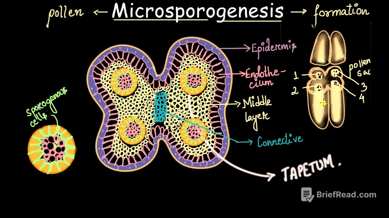
The video describes the structure of the anther, focusing on a bilobed (dithicus) anther, which has two lobes and two compartments. In a transverse section of the anther, tube-like structures called pollen sacs are visible, where pollen grains are produced. Each lobe contains two pollen sacs, resulting in a total of four pollen sacs in a dithicus anther. The number of pollen sacs is emphasized due to its relevance in exam questions.
Layers of the Anther Wall [3:23]
Zooming in on the anther, the outermost layer is the epidermis, a single sheet of cells. The two lobes of the anther are connected by a tissue called the connective, through which vascular tissue provides nourishment. Beneath the epidermis is the endothecium, and further inside is the middle layer. The epidermis, endothecium, and middle layer collectively provide protection to the developing microspores. Inside these layers, pink cells are visible, which will eventually develop into microspores. The most important layer is the tapetum, which surrounds these cells and has several functions.
Tapetum and Sporogenous Cells [5:15]
The video provides a closer look at the tapetum layer and the cells inside. The tapetal cells are large, contain a lot of cytoplasm, and provide nourishment to the inner cells. The tightly packed cells inside the tapetum are called sporogenous cells, which will develop into microspores. Not all sporogenous cells develop into microspores; some disintegrate and are consumed by neighboring cells, acting as a food source. The remaining cells develop into microspores or pollen grains.
Microspore Mother Cell and Meiosis [6:49]
One of the remaining sporogenous cells, now called a microspore mother cell, is examined. Plants exist in either the gametophyte (haploid) or sporophyte (diploid) stage. To produce pollen, which is a male gamete with a haploid number of chromosomes, the microspore mother cell undergoes meiosis. The microspore mother cell, being in the sporophyte stage, has a diploid number of chromosomes (2n).
Microspore Tetrad Formation
The microspore mother cell undergoes meiosis, resulting in four haploid cells. These four cells are collectively called a microspore tetrad. The arrangement of these cells can vary, including tetrahedral, linear, iso-bilateral, and T-shaped arrangements, with the tetrahedral shape being the most common. The microspore tetrad is held together by a protein called callose.
Release of Microspores and Microsporogenesis
Each of the four haploid cells in the tetrad develops into individual pollen particles. To separate these cells, the callose layer needs to be dissolved. The tapetum layer produces an enzyme called callase, which dissolves the callose, releasing the four individual microspores. This entire process, from the microspore mother cell to the formation of individual microspores, is called microsporogenesis. The video clarifies that these microspores are not yet fully developed pollen grains and require further changes.
Calculating Pollen Count
The video emphasizes that one microspore mother cell yields four microspores, which will become four pollen grains. If a pollen sac contains five microspore mother cells, it will produce 20 pollen grains (5 x 4). In a dithicus anther, which has four pollen sacs, each containing five microspore mother cells, a total of 80 pollen grains will be produced (4 x 20). This explains the abundance of pollen in flowers. The video concludes by mentioning that future videos will cover the changes that occur in these individual cells to form fully developed pollen grains.