TLDR;
This video provides a detailed anatomical overview of a pig heart, focusing on both external and internal structures. It covers how to orient the heart, identify key chambers and valves, and trace the major blood vessels connected to the heart. The tutorial uses clear explanations and visual aids to help viewers understand the complex structure and function of the heart.
- Orientation and External Anatomy: The video begins by showing how to orient the heart, identifying the base and apex, and locating the coronary artery.
- Internal Anatomy and Chambers: The tutorial explains how to identify the left and right ventricles and atria, along with their respective valves (bicuspid and tricuspid).
- Blood Vessels: The video details the major blood vessels, including the aorta, pulmonary artery, vena cava, and pulmonary vein, explaining which chambers they connect to and their functions.
External Anatomy and Orientation [0:00]
The pig heart is oriented with the top part, or base, where major blood vessels enter and exit. The bottom part, which comes to a point, is known as the apex. The coronary artery, which feeds the heart, runs down around the heart and branches off. This artery is one of the first to branch off the aorta, the main blood vessel from the heart.
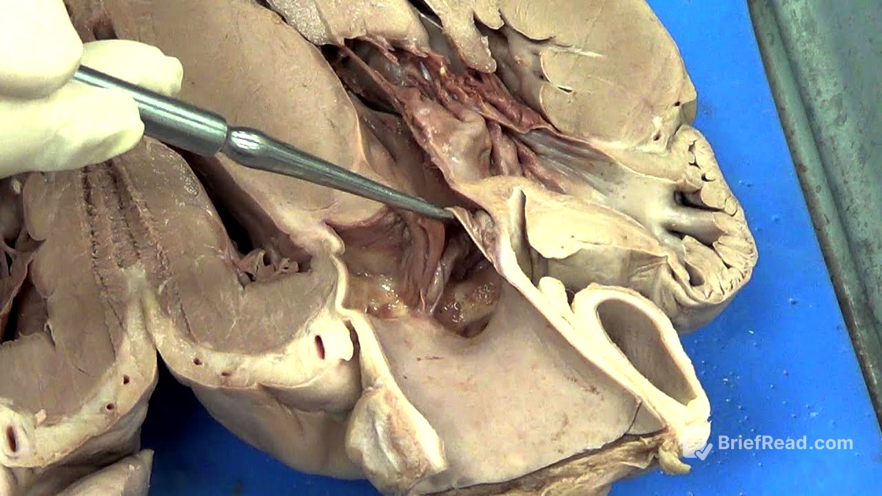
Internal Anatomy and Chambers [0:52]
The heart is bisected to reveal its internal chambers. The left ventricle is identified as the thickest, most muscular chamber, responsible for pumping blood to the entire body. The bicuspid valve leads to the left ventricle, featuring chordae tendineae attached to papillary muscles that control valve opening. Above the bicuspid valve is the left atrium. The right ventricle, less muscular than the left, also has a valve, the tricuspid valve, leading to it. Above the tricuspid valve is the right atrium, which connects to an extension called the auricle.
Major Blood Vessels [4:23]
There are four major blood vessels associated with the heart: two veins entering the atria and two arteries exiting the ventricles. Arteries, which carry blood away from the heart, have thick, strong walls to withstand high pressure. The aorta, the largest artery, exits the left ventricle, separated by the aortic semilunar valve. The pulmonary artery exits the right ventricle. Thin-walled holes indicate veins.
Veins and Atria [7:18]
Thin-walled blood vessels that collapse when released are likely veins. A probe inserted into a large hole reveals it enters the right atrium, identifying it as the vena cava. The vena cava can sometimes have one or two holes (superior and inferior). Another large, thin-walled vessel entering the left atrium is the pulmonary vein. Additional holes near the pulmonary artery are branches of it.
Coronary Artery and Aorta [10:11]
Small holes observed are likely branches of the coronary artery. The first blood vessel coming off the aorta leads to the coronary artery, which feeds the heart. A probe inserted into a small hole near the aorta confirms its connection to the coronary artery.