TLDR;
This video provides a detailed explanation of the neural and sensory systems, focusing on their structure, function, and coordination in maintaining homeostasis. It covers the nervous system's organization, neuron types, nerve impulse conduction, synaptic transmission, brain structure, reflex actions, eye anatomy and function, and ear anatomy and function.
- Neural and endocrine systems maintain homeostasis.
- Neurons are structural and functional units of the neural system.
- The human eye functions like a camera, using photoreceptors to generate impulses.
- The ear is responsible for hearing and maintaining body balance.
Coordination and Integration [0:00]
Coordination between organs and systems is essential for maintaining homeostasis. Different organs form systems with specialized functions that interact and depend on each other. Physical exercise exemplifies this, where increased energy demand leads to a higher respiration rate, heartbeat, and blood flow to muscles, along with waste expulsion and temperature regulation. The neural and endocrine systems maintain homeostasis through quick nerve impulses and chemical integration via hormones.
Neural System Organization [2:06]
All animals possess a neural system composed of neurons, which perceive, receive, and transmit stimuli. Neural organization varies across species. Sponges lack neurons, while Hydra has a network. Flatworms have a ladder-type system with nerve rings and cords. Earthworms feature a circumferential nerve ring and a ganglionated nerve cord. Insects have a more organized system, and starfish have nerves connected by a central ring. Vertebrates have well-developed neural systems, with primates, especially humans, having the most complex brains.
Human Neural System [3:40]
The human neural system includes the central neural system (CNS) and the peripheral neural system (PNS). The CNS, consisting of the brain and spinal cord, processes information. The PNS comprises cranial and spinal nerves, controlling voluntary functions. Peripheral nerves are afferent (sensory) or efferent (motor). Afferent fibers carry impulses from sense organs to the CNS, while efferent fibers carry impulses from the CNS to involuntary organs, muscles, and glands. The PNS divides into the somatic and autonomic neural systems. The somatic system relays impulses to skeletal muscles, and the autonomic system controls involuntary functions like heartbeat and peristalsis. The autonomic system is further classified into sympathetic and parasympathetic systems, originating from thoracic-lumbar and cranial-sacral regions, respectively.
Neuron Structure and Types [5:42]
A neuron, the longest cell in the body, consists of the cyton and nerve processes. The cyton, or soma, contains granular cytoplasm (neuroplasm), a nucleus, Nissl granules for protein synthesis, mitochondria for energy, and neurofibers for impulse conduction. Processes include dendrites and axons. Dendrites, which can be multiple, transmit impulses toward the cell body. An axon, always single, is covered by the axolemma, with inner axoplasm. The distal end forms a synaptic knob containing synaptic vesicles filled with neurotransmitters. Neurons are classified into multipolar (one axon, multiple dendrites), bipolar (one axon, one dendrite), and unipolar (one axon only). Nerve fibers are either myelinated or non-myelinated. Myelinated fibers have Schwann cells forming a myelin sheath with nodes of Ranvier, found in cranial and spinal nerves. Unmyelinated fibers are enclosed by Schwann cells without forming a myelin sheath, found in autonomic and somatic systems. Based on function, neurons are sensory (afferent), motor (efferent), or association (interneurons). Sensory neurons receive impulses from receptors and direct them to the CNS. Motor neurons carry impulses from the CNS to organs. Association neurons interlink sensory and motor neurons within the CNS.
Nerve Impulse Conduction [10:07]
A nerve impulse is a mechanical, chemical, and electrical disturbance caused by a stimulus in a neuron. Conduction involves resting membrane potential and action membrane potential. Neurons are excitable cells stimulated by physical, chemical, or electrical stimuli. The nerve fiber is covered by a neural membrane with voltage-gated sodium and potassium ion channels. In a resting membrane, these channels are differentially permeable, more so to potassium ions and less to sodium ions. The axoplasm contains high potassium and low sodium concentrations, while the extracellular fluid has low potassium and high sodium concentrations. This is maintained by a sodium-potassium pump, which transports three sodium ions out for every two potassium ions in, creating an electrical potential difference called resting potential. When a stimulus is applied, sodium channels open, causing rapid influx of sodium ions and reversing the polarity (depolarization). This creates an action potential that travels as a wave along the nerve fiber.
Action Potential and Synapse [13:52]
The action potential travels along the nerve fiber as a wave of depolarization. At site B, the axon membrane has a positive charge on the outer surface and a negative charge on the inner surface. Current flows from site A to site B on the inner surface and from site B to site A on the outer surface, reversing the polarity at site B and generating an action potential. The sequence repeats along the axon. At the peak of action potential, sodium permeability decreases, and potassium permeability increases, leading to repolarization. The membrane regains its original polarity within milliseconds. A repolarized nerve fiber undergoes a refractory period, where the sodium-potassium pump restores the original ionic distribution. A synapse transmits the nerve impulse from one neuron to another, formed by the membranes of pre- and postsynaptic neurons, separated by a synaptic cleft.
Synaptic Transmission [16:01]
Synapses are either chemical or electrical. Chemical synapses, common in humans, consist of a presynaptic neuron, synaptic cleft, and postsynaptic neuron. The presynaptic neuron ends in a synaptic knob with mitochondria and synaptic vesicles containing neurotransmitters like acetylcholine. The synaptic cleft is a fluid-filled gap with no protoplasmic continuity. When an action potential arrives, synaptic vesicles move towards the presynaptic membrane, fuse, and release neurotransmitters into the synaptic cleft. These neurotransmitters bind to chemo receptors on the postsynaptic membrane, opening sodium ion channels and generating a new potential, which may be excitatory or inhibitory. Electrical synapses, less common, feature pre- and postsynaptic neurons in close proximity, allowing direct electric current flow. Synaptic vesicles are absent, and few mitochondria are present. Impulse transmission is faster in electrical synapses than in chemical synapses.
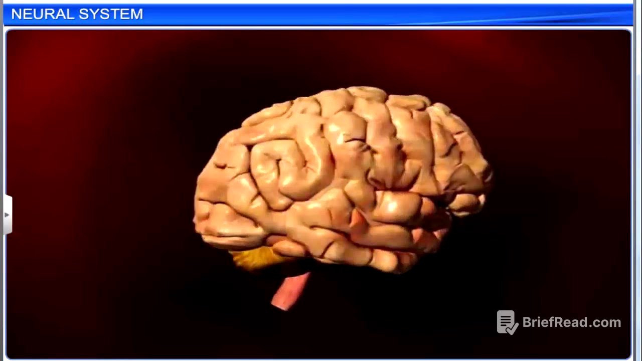
Brain Structure and Function [18:50]
The brain and spinal cord form the central neural system. The brain, the central information processing organ, controls voluntary and involuntary movements, vision, hearing, speech, memory, intelligence, emotions, and thought. It is protected by the skull and three meninges: dura mater, arachnoid, and pia mater. The space between the arachnoid and pia mater is filled with cerebrospinal fluid, which acts as a shock absorber. The brain is divided into the forebrain, midbrain, and hindbrain. The forebrain includes the cerebrum, thalamus, and hypothalamus. The cerebrum is divided into left and right hemispheres connected by the corpus callosum. The cerebral cortex, or gray matter, contains motor, sensory, and association areas. The inner part of the hemisphere is white matter due to myelinated axons. The thalamus coordinates sensory and motor signaling. The hypothalamus controls body temperature, circadian rhythms, and urges to eat and drink, and it secretes hormones that control the pituitary gland. The limbic system, including the amygdala and hippocampus, regulates sexual behavior, long-term memory, olfaction, and emotional reactions.
Midbrain and Hindbrain [23:22]
The midbrain, located between the forebrain and hindbrain, contains the cerebral aqueduct. The dorsal portion has four lobes called corpora quadrigemina, which contain reflex centers for vision and hearing. The midbrain and hindbrain form the brainstem, a relay station for auditory and visual information. The hindbrain consists of the pons, cerebellum, and medulla oblongata. The pons interconnects the cerebral cortex and medulla to the cerebellum. The cerebellum coordinates body movement and balance. The medulla oblongata links the brain and spinal cord and controls involuntary activities like respiration, blood pressure, cardiovascular reflexes, and gastric secretions.
Reflex Action [25:39]
Reflex action is a quick, involuntary response to an external stimulus without conscious thought, involving the spinal cord. The reflex arc includes a receptor, sensory neuron, interneuron, motor neuron, and effector organ. The afferent neuron receives a nerve impulse from sensory organs and transmits it through the dorsal nerve root to the spinal cord. The sensory neuron connects to an association neuron, which transmits the stimulus to the effector neuron. The efferent neuron passes the stimulus to effector organs like muscles through the ventral nerve cord. The reflex arc saves time by bypassing the brain, helping avoid painful experiences.
Eye Anatomy and Function [27:16]
The eye detects environmental changes and sends signals to the CNS. Human beings have spherical eyes in eye sockets, adapted for binocular vision, protected by eyebrows, eyelids, and lacrimal glands. The eye wall has three layers: the sclera (external), choroid (middle), and retina (inner). The sclera's anterior portion is the cornea. The choroid contains blood vessels and forms the ciliary body and iris. The lens is held by ligaments of the ciliary body. The iris surrounds the pupil, regulating its diameter. The retina contains ganglion cells, bipolar cells, and photoreceptor cells (rods and cones). Cones are responsible for color and daylight vision (photopic), while rods are responsible for twilight vision (scotopic). Rhodopsin, a purple pigment, is present in rods and contains a derivative of vitamin A. Cones are of three types, responding to red, green, and blue light. The retina has a blind spot where the optic nerve leaves and a fovea with high cone density for best visual acuity.
Visual Process [31:51]
The aqueous chamber, filled with aqueous humor, lies between the cornea and lens, providing nutrition. The vitreous chamber, filled with vitreous humor, lies between the lens and retina, maintaining eye shape and intraocular pressure. The eye functions like a camera, with the cornea, aqueous humor, lens, and vitreous humor refracting light rays to focus on the retina. The iris acts as a diaphragm, regulating light entry. Light stimulates photoreceptors, forming an inverted image. The brain interprets nerve impulses, perceiving the image correctly. Light dissociates retinal from opsin, changing opsin's structure and membrane permeability, resulting in potential differences in photoreceptor cells. This generates action potentials in ganglion cells through bipolar cells. Impulses are transmitted to the visual area of the cerebral cortex via the optic nerve, where they are analyzed to recognize the image.
Ear Anatomy [34:02]
The ear assists in hearing and maintaining balance, divided into the outer, middle, and inner ear. The outer and middle ear structures persist only in hearing, while the inner ear helps maintain equilibrium. The outer ear includes the pinna and external auditory meatus, which has hairs and wax-secreting glands. The pinna collects vibrations and directs sound into the meatus. The middle ear consists of the tympanic cavity, separated from the outer ear by the tympanic membrane (eardrum) and from the inner ear by an auditory capsule with oval and round windows. The middle ear contains ossicles (malleus, incus, stapes) that increase the efficiency of sound wave transmission. The Eustachian tube connects the tympanic cavity to the pharynx, maintaining air pressure. The inner ear consists of the labyrinth, with the cochlea for hearing and the vestibular apparatus for balance. The bony labyrinth is filled with perilymph, while the membranous labyrinth is filled with endolymph.
Inner Ear Structure and Function [37:56]
The cochlea, the coiled portion of the bony labyrinth, contains Reissner's membrane and the basilar membrane, dividing the labyrinth into the scala vestibuli and scala tympani, both filled with perilymph. The space between these is the scala media, filled with endolymph. The organ of Corti, located in the scala media, is the receptor of sound, composed of hair cells that act as auditory receptors. Above the hair cells lies the tectorial membrane. The vestibular apparatus, located above the cochlea, maintains balance and consists of semicircular canals and the otolith organ (saccule and utricle). The semicircular canals contain a sensory spot called the crista ampullaris, while the saccule and utricle contain a sensory spot called the macula. Both crista and macula contain hair cells that act as receptors for balance and posture.
Hearing Process [40:55]
The external ear receives sound waves and directs them to the eardrum. The eardrum vibrates and transmits the vibrations through the ossicles to the oval window. The ossicles amplify the sound and pass the vibrations to the fluid of the cochlea, generating waves in the lymph that induce a ripple in the basilar membrane. The vibrations of the basilar membrane bend the hair cells against the tectorial membrane, stimulating the hair cells. The hair cells generate nerve impulses, which are transmitted along afferent nerve fibers and auditory nerves to the auditory cortex of the brain. The brain interprets these nerve impulses, and sound is recognized. The ear forms an important part of the auditory system and helps maintain balance.