TLDR;
This video provides an overview of the thoracic wall, including its bony framework, openings, and internal cavities. It details the structures forming the superior and inferior thoracic apertures, the components of the bony thorax (vertebrae, ribs, and sternum), and the three internal cavities: two pleural cavities for the lungs and the mediastinum for the heart, major vessels, and other structures. The video also explains the classification of ribs into true, false, and floating ribs based on their attachment to the sternum.
- The thoracic wall extends from the superior to the inferior thoracic aperture, protecting vital structures.
- The bony framework consists of thoracic vertebrae, ribs, and the sternum.
- The thoracic cavity contains two pleural cavities (lungs) and the mediastinum (heart, major vessels).
- Ribs are classified as true (1-7), false (8-12), and floating (11-12) based on their sternal attachments.
Introduction to the Thoracic Wall [0:00]
The video introduces the thoracic wall, which extends from the superior thoracic aperture to the inferior thoracic aperture. The thoracic wall is composed of a bony framework that protects the vital structures within the thoracic cavity. The video will cover the thoracic apertures, the bony framework, and the internal cavities of the thorax.
Superior Thoracic Aperture [0:22]
The superior thoracic aperture is formed posteriorly by the body of the first thoracic vertebra (T1). Laterally, it is bordered by the medial margin of the first rib. Anteriorly, the manubrium of the sternum completes the aperture. Structures such as vessels and nerves pass through this opening to and from the head, neck, and upper limbs.
Inferior Thoracic Aperture [1:04]
The inferior thoracic aperture is defined by several structures. Anteriorly, it is marked by the costal margin, while laterally, it includes the distal end of the 11th rib and the entire 12th rib. Posteriorly, the body of the 12th thoracic vertebra (T12) forms the boundary. This aperture is largely closed by the diaphragm muscle, through which structures like the aorta, inferior vena cava, and esophagus pass.
Bony Framework of the Thorax [2:32]
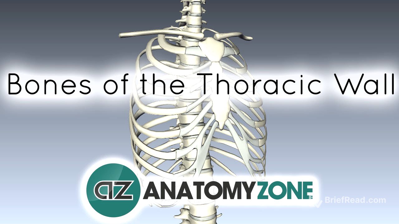
The bony framework of the thorax consists of the 12 thoracic vertebrae posteriorly, the 12 pairs of ribs laterally, and the sternum anteriorly. The ribs connect to the thoracic vertebrae and articulate with the sternum via costal cartilages. The sternum is composed of three parts: the manubrium, the body, and the xiphoid process.
Internal Cavities of the Thorax [3:14]
The thorax contains three main cavities: two pleural cavities and the mediastinum. The pleural cavities, located laterally, house the lungs. The mediastinum, situated between the pleural cavities, contains the heart, major blood vessels, trachea, esophagus, various nerves, and lymphatic structures. The thoracic wall protects the vital structures within these cavities.
Rib Classification [4:01]
There are 12 pairs of ribs that articulate with the 12 thoracic vertebrae. These ribs are classified into true, false, and floating ribs. The first seven ribs are true ribs because their costal cartilages directly articulate with the sternum. Ribs 8-12 are false ribs; their costal cartilages do not directly articulate with the sternum but connect to the cartilage of the rib above. Ribs 11 and 12 are floating ribs because they do not have any anterior attachment to the sternum or other costal cartilages.


