TLDR;
This video provides a detailed explanation of the bony features of the radius, a lateral bone of the forearm. It covers the upper end, shaft, and lower end of the radius, describing their key anatomical landmarks, surfaces, and articulations.
- The upper end includes the head, neck, and radial tuberosity, which contribute to the elbow and superior radioulnar joints.
- The shaft has three borders (anterior, posterior, and interosseous) and three surfaces (anterior, posterior, and lateral), each with distinct features like the nutrient foramen and oblique lines.
- The lower end is expanded with five surfaces (anterior, posterior, medial, lateral, and inferior), featuring the ulnar notch, styloid process, and articular surfaces for the wrist joint.
Introduction to the Radius [0:00]
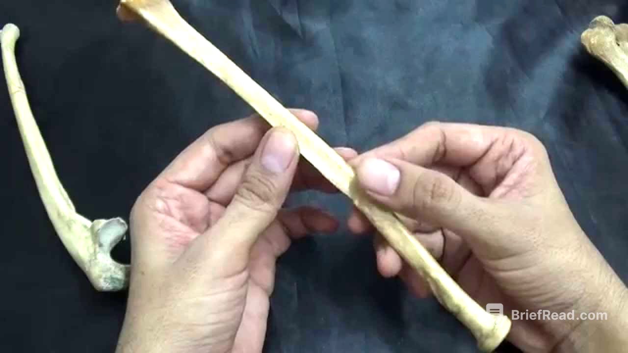
The radius is a lateral bone of the forearm, homologous to the tibia in the lower limb. As a typical long bone, it consists of an upper end, a lower end, and a shaft. The shaft features three borders and three surfaces, along with a nutrient foramen.
Upper End of the Radius [0:23]
The upper end of the radius includes the head, which is disc-shaped and covered with hyaline cartilage. The superior concave surface of the head articulates with the capitulum of the humerus, forming part of the elbow joint (humero-radial joint). The circumference of the head articulates with the radial notch of the ulna and the annular ligament, forming the superior radioulnar joint, a part of the radio-synovial joint. The constricted neck is partly enclosed by the lower margin of the annular ligament. Next to the neck is the radial tuberosity, which is rough posteriorly and smooth anteriorly.
Shaft of the Radius: Borders [2:52]
The shaft of the radius has three borders: anterior, posterior, and medial (interosseous). The anterior border extends from the anterior margin of the radial tuberosity to the styloid process. Its upper half is oblique (anterior oblique line), while the lower half is vertical and crest-like. The posterior border, a mirror image of the anterior border, is most visible in the middle third of the shaft. It also features a posterior oblique line in its upper half. The medial border, or interosseous border, is the sharpest and provides attachment for the interosseous membrane. It extends from the radial tuberosity above to the posterior margin of the ulnar notch below.
Shaft of the Radius: Surfaces [5:47]
The radius has three surfaces: anterior, posterior, and lateral. The anterior surface lies between the anterior and interosseous borders and contains a nutrient foramen directed upwards, supplied by a branch of the anterior interosseous artery. The posterior surface lies between the posterior and medial (interosseous) borders. The lateral surface is located between the anterior and posterior borders and is rough in the middle third.
Lower End of the Radius [6:57]
The lower end of the radius is expanded and has five surfaces: anterior, medial, lateral, posterior, and inferior. The anterior surface is a continuation of the shaft's anterior surface and is relatively smooth. The posterior surface has three to four grooves and the tubercle of Lister, which is lateral to an oblique groove lodging the tendon of extensor pollicis longus. The medial surface features the ulnar notch for articulation with the head of the ulna, forming the inferior radioulnar joint. The lateral surface is prolonged to form the styloid process. The inferior surface is articular, smooth, and covered by hyaline cartilage, with a triangular articular surface laterally for the scaphoid and a quadrangular articular surface medially for the lunate bone. These bones, along with the articular disc, form the wrist joint or radiocarpal joint.


![BEST MACD Trading Strategy [86% Win Rate]](https://wm-img.halpindev.com/p-briefread_c-10_b-10/urlb/aHR0cDovL2ltZy55b3V0dWJlLmNvbS92aS9yZl9FUXZ1Yktsay9ocWRlZmF1bHQuanBn.jpg)