TLDR;
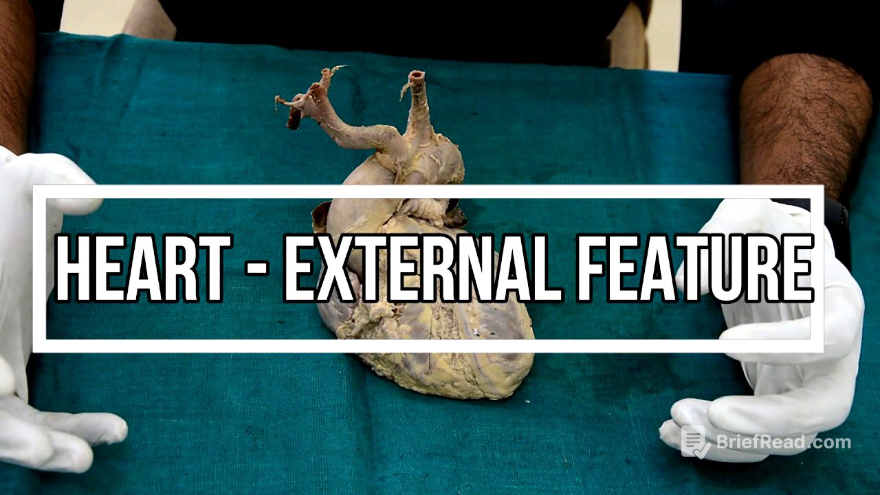
This video provides an overview of the external features of the heart, including its location, shape, size, and various surfaces and borders. It also explains the anatomical position of the heart for examination purposes.
- The heart is located in the middle mediastinum, within the pericardium in the thoracic cavity.
- The heart has a conical or pyramidal shape, with an average size of 12 by 9 centimetres.
- Key external features include the apex, base, borders (right, left, inferior, superior), and surfaces (sternocostal, left, diaphragmatic).
Introduction to the Heart [0:09]
The heart, a hollow muscular organ, resides in the middle mediastinum within the pericardium in the thoracic cavity. Known as "cardia" in Greek and "cordis" in Latin, the heart has a conical or pyramidal shape, averaging 12 by 9 centimetres in size, slightly larger than one's clenched fist. Weighing about 300 grams in males and 250 grams in females, the heart is positioned behind the sternum, with one-third lying to the right and two-thirds to the left of the median plane. It comprises four chambers: two atria and two ventricles. The atria are located upwards and backwards, while the ventricles are downward and forward. The right atrium receives the superior and inferior vena cava, and the left atrium receives four pulmonary veins.
External Features: Apex and Base [2:14]
The heart has an apex, a base, borders (right, left, inferior and superior), and surfaces (sternocostal, left and diaphragmatic). The apex, formed by the left ventricle, points downward, forward, and to the left, located in the left fifth intercostal space, nine centimetres from the midline, inside the midclavicular line. Conversely, the base faces upward, backward, and to the right. It is quadrilateral in shape, formed primarily by the left atrium (two-thirds) and partly by the right atrium (one-third).
Borders of the Heart [3:56]
The heart has four borders: right, left, inferior, and superior. The right border, formed by the right atrium, is rounded and convex, extending from the superior vena cava to the inferior vena cava. A shallow vertical groove, the sinus terminalis, is visible on the surface. The left border, convex and facing upward and to the left, extends from the left auricle to the apex, accompanied by the marginal branch of the left coronary artery. The inferior border is horizontal, extending from the inferior vena cava opening to the apex, mainly formed by the right ventricle and partly by the right atrium, accompanied by the right marginal branch of the right coronary artery. Near the apex, the inferior border presents the incisura cordis and separates the sternocostal surface from the diaphragmatic surface. The superior border is slightly oblique and obscured anteriorly by the aorta and pulmonary trunk, mainly formed by the left atrium and partly by the right atrium.
Surfaces of the Heart [6:31]
The heart has three surfaces: sternocostal, left, and diaphragmatic. The sternocostal surface, large and convex, faces upwards and forwards, mainly formed by the right ventricle, right atrium, and auricle, and partly by the left ventricle and left auricle. It features the anterior part of the right atrioventricular groove and the anterior interventricular groove, which houses the anterior interventricular artery. The diaphragmatic surface is flat, with its left two-thirds formed by the left ventricle and the right one-third by the right ventricle. It contains the posterior interventricular groove, lodging the posterior interventricular artery. The left surface faces upwards, backwards, and to the left, mainly formed by the left ventricle and partly by the left atrium and left auricle.
Grooves and Sulci of the Heart [8:27]
The heart features several grooves or sulci. The atrioventricular groove (coronary sulcus) separates the atria from the ventricles in a C-shape, deficient anteriorly due to the pulmonary trunk. It has anterior and posterior parts; the anterior part is divided into right and left halves. The right half contains the trunk of the right coronary artery, while the left half contains the circumflex branch of the left coronary artery, the termination of the great cardiac vein, and the commencement of the coronary sinus. The posterior atrioventricular groove contains only the coronary sinus. The interventricular sulcus separates the two ventricles, with anterior and posterior portions. The anterior interventricular groove, on the sternocostal surface, lodges the anterior interventricular artery (a branch of the left coronary artery). The posterior interventricular groove, on the diaphragmatic surface, lodges the posterior interventricular artery (mostly a branch of the right coronary artery). The interatrial groove, separating the two atria, is deficient anteriorly due to the aorta and pulmonary trunk and faint posteriorly. The meeting point of the interatrial groove, coronary sulcus, and posterior interventricular groove is known as the crux of the heart.
Key Features and Anatomical Position [11:31]
The apex is formed only by the left ventricle, the base by the left and right atria, the diaphragmatic surface by the right and left ventricles, and the right border by the right atrium. Two-thirds of the heart lie to the left of the midline, while one-third lies to the right. Two-thirds of the sternocostal surface are formed by the right ventricle, and one-third by the left ventricle. Conversely, two-thirds of the diaphragmatic surface are formed by the left ventricle, and one-third by the right ventricle. Two-thirds of the base are formed by the left atrium, and one-third by the right atrium. To maintain the anatomical position during examination, the apex should point downward, forward, and to the left, the inferior border should lie horizontally, and the flat diaphragmatic surface should rest on the palm of the left hand.


