TLDR;
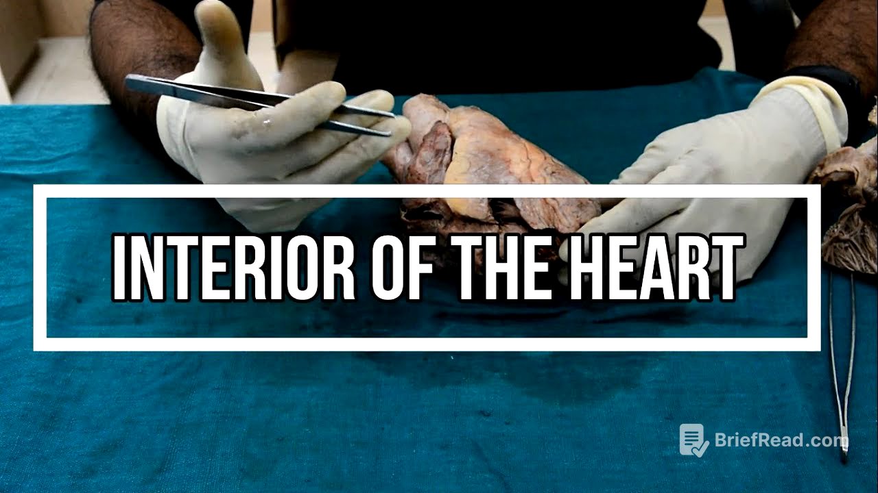
This video provides a detailed overview of the interior of the heart, focusing on each of the four chambers: the right atrium, left atrium, right ventricle, and left ventricle. It explains the key anatomical features, including the openings of major blood vessels, the structure of the valves, and the muscular components of each chamber. The video also touches on the embryological origins of certain structures and their functional significance.
- The right atrium receives deoxygenated blood from the superior and inferior vena cava and the coronary sinus.
- The left atrium receives oxygenated blood from the four pulmonary veins.
- The right ventricle pumps deoxygenated blood to the pulmonary trunk.
- The left ventricle pumps oxygenated blood to the aorta.
Introduction [0:09]
The video introduces a discussion on the interior of the heart, following a previous video that covered the external features. The heart is composed of four chambers: two atria (right and left) and two ventricles (right and left). The video aims to explore the interior of each of these chambers in detail.
Right Atrium [0:59]
The right atrium forms part of the sternocostal surface, the right border, and part of the upper border of the heart. It receives deoxygenated blood from the entire body via the superior and inferior vena cava. The right atrium is elongated, with the superior vena cava opening at its upper end and the inferior vena cava at its lower end. The right auricle, an elongated extension of the upper end, has a spongy interior to prevent free blood flow. The sulcus terminalis, located externally along the right border, corresponds to the crista terminalis internally. The sinoatrial node, which acts as the heart's pacemaker, is situated in the upper part of the sulcus terminalis. The right atrium is separated from the right ventricle by the right atrioventricular groove, which houses the right coronary artery and small cardiac vein.
Interior of the Right Atrium [3:16]
The interior of the right atrium features a smooth posterior part (sinus venarum) and a rough anterior part (atrium proper), separated by the crista terminalis. The sinus venarum develops from the absorbed right horn of the sinus venosus, while the atrium proper develops from the right half of the primitive atrium. The sinus venarum contains the openings of the inferior vena cava, superior vena cava, and coronary sinus. A web-like network, known as the Chiari network, may sometimes be present near the opening of the inferior vena cava. The posterior wall of the sinus venarum also contains the opening of the foramen venarum minimum. The rough anterior part shows transverse muscular ridges called musculi pectinati, which arise from the crista terminalis and extend towards the right atrioventricular orifice.
Interatrial Septum [6:06]
The interatrial septum, located between the right and left atria, develops from the septum primum and septum secundum. It features the fossa ovalis, a saucer-shaped depression, and the limbus fossa ovalis (annulus ovalis), a prominent margin developed from the septum secundum. Remnants of the foramen ovale may sometimes be present between the fossa ovalis and the limbus.
Left Atrium [7:04]
The left atrium is situated posterior and to the left of the right atrium. It forms the left two-thirds of the base and the upper border of the heart. Anterosuperiorly, it features the left auricle, a conical projection. The left atrium receives oxygenated blood from the four pulmonary veins.
Interior of the Left Atrium [8:04]
The interior of the left atrium is derived embryologically from the absorbed pulmonary veins. Most of its interior is smooth, except for the left auricle, which has a rough, muscular pattern developed from the original primitive atrium. The interatrial septum in the left atrium shows the impression of the limbus fossa ovalis, corresponding to the fossa ovalis in the right atrium.
Right Ventricle [9:55]
The right ventricle is triangular in shape and forms the right one-third of the diaphragmatic surface and the wall of the inferior border. Its interior is divided into a lower rough (inflowing) part and a smooth upper (outflowing) part. The rough part features trabeculae carneae, muscular ridges of three types: ridges, bridges, and papillary muscles. Papillary muscles attach to the cusps of the tricuspid valve via chordae tendineae. There are three papillary muscles in the right ventricle: anterior (largest), posterior (smallest), and septal (small, numerous). The base of the anterior papillary muscle is attached to the interventricular septum via the septomarginal trabecula (moderator band), which prevents overdistension of the right ventricle. The interventricular septum bulges towards the right ventricle, giving its cavity a crescent shape. The wall of the right ventricle is thinner than that of the left ventricle, with a thickness ratio of 1:3.
Smooth Upper Part of Right Ventricle [14:28]
The smooth upper part of the right ventricle is conical and gives rise to the pulmonary trunk. This outflowing part is separated from the rough lower part by the supraventricular crest (infundibulo-ventricular crest), which lies between the pulmonary orifice and the right atrioventricular orifice.
Interventricular Septum [15:15]
The interventricular septum lies obliquely, with its lower part facing forward and to the right, and its upper part facing backward and to the left. It separates the two ventricles, and its upper thin part separates the left ventricle from the right atrium. The position of the interventricular septum on the surface is indicated by the anterior and posterior interventricular grooves.
Left Ventricle [15:56]
The left ventricle is conical in shape and forms the left one-third of the sternocostal surface, the left two-thirds of the diaphragmatic surface, the apex, and the left border of the heart. Similar to the right ventricle, it has a rough (inflowing) part with trabeculae carneae and a smooth (outflowing) part. The inflowing part develops from the primitive ventricle of the heart tube. Unlike the right ventricle, it has only two papillary muscles: anterior and posterior, which attach to the cusps of the mitral valve via chordae tendineae.
Outflowing Part of Left Ventricle [17:01]
The outflowing part of the left ventricle, known as the aortic vestibule, leads into the aortic orifice, which is guarded by the aortic valve. The aortic vestibule develops from the mid-portion of the bulbus cordis. The left ventricle features the left atrioventricular orifice (bicuspid or mitral orifice), guarded by the mitral valve, and the aortic orifice, guarded by the semilunar aortic valve.
Orifices and Valves [18:09]
The video concludes by summarising the orifices and valves connecting the different chambers of the heart. The right atrium connects to the right ventricle through the right atrioventricular orifice (tricuspid orifice), guarded by the tricuspid valve. Blood from the right ventricle is pushed to the pulmonary trunk through the pulmonary orifice, guarded by the semilunar pulmonary valve. Blood enters the left atrium via the pulmonary veins and passes to the left ventricle through the left atrioventricular orifice, guarded by the mitral valve (bicuspid valve). Finally, blood from the left ventricle passes to the ascending aorta through the aortic opening, guarded by the semilunar aortic valve.


