TLDR;
This text describes computed tomography (CT), a non-invasive imaging technique that uses X-rays and computer analysis to create detailed cross-sectional images of the body. CT scanning is valuable for visualizing soft tissues and measuring structures with millimeter precision, but it exposes patients to higher radiation doses compared to traditional X-rays, posing an increased cancer risk, especially for children and those undergoing multiple scans.
- CT uses X-rays to create detailed cross-sectional images.
- It is particularly useful for soft tissue scanning.
- CT scans expose patients to higher radiation doses, increasing cancer risk.
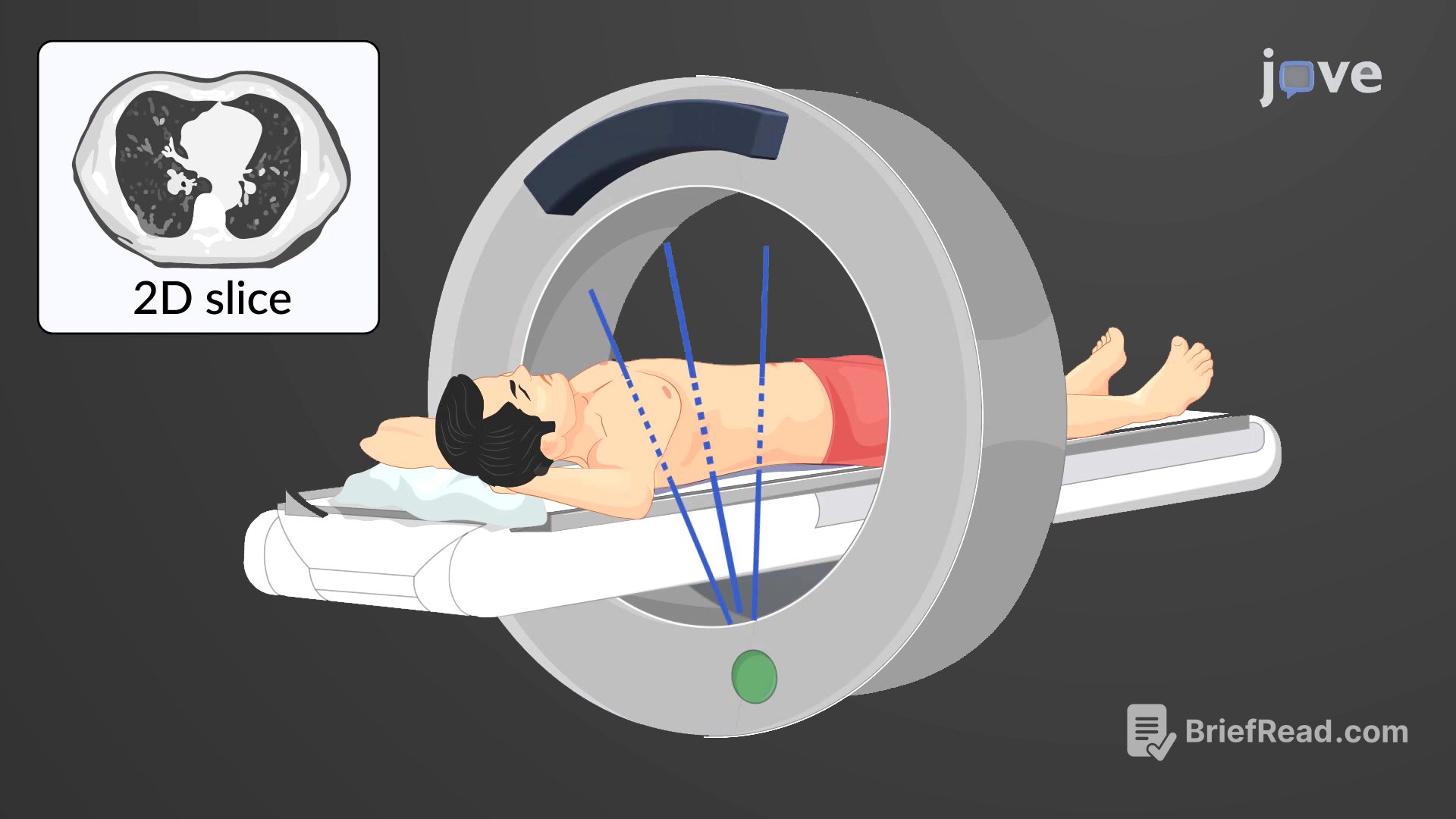
Computed Tomography
Computed tomography (CT) is a non-invasive imaging method that uses computers to analyze multiple cross-sectional X-rays, providing detailed views of internal body structures. Invented in the 1970s, CT scanning works by measuring the absorption and reflection of X-rays as they pass through the body. During a CT scan, the patient lies on a motorized platform while a computerized axial tomography scanner rotates 360 degrees, capturing X-ray images from all angles. These images are then combined by a computer to create a two-dimensional "slice" of the scanned area. Modern advancements in computer technology and software have made CT scanning a routine diagnostic tool, especially useful for examining soft tissues in the brain, thorax, and abdominal viscera. The precision of CT scans allows physicians to measure tumors and tissues down to the millimeter. Furthermore, software can reformat the image slices to view different anatomical planes, such as sagittal and coronal, reducing the need for additional X-ray exposure. However, CT scanning exposes patients to significantly higher doses of radiation compared to traditional X-rays, increasing the risk of cancer, particularly in children and adults who undergo multiple scans.