TLDR;
This grand rounds lecture by Dr. Yuichi Shimada focuses on hypertrophic cardiomyopathy (HCM), covering its introduction, diagnosis, and management. The lecture emphasizes HCM as a significant cause of sudden cardiac death in young athletes, discusses the genetic and phenotypic variations, and provides guidance on diagnostic approaches, including ECG and echocardiography. It also addresses treatment strategies, including activity restrictions, family screening, prevention of sudden cardiac death with ICDs, and symptom management with medications and invasive procedures.
- HCM is a leading cause of sudden cardiac death in young athletes.
- Genetic testing has limitations due to numerous genes and mutations.
- Diagnosis involves recognizing LV hypertrophy without other causes.
- Management includes activity restrictions, family screening, and ICD implantation.
- Symptom control focuses on reducing LVOT obstruction with medications or surgery.
Introduction to Hypertrophic Cardiomyopathy [2:44]
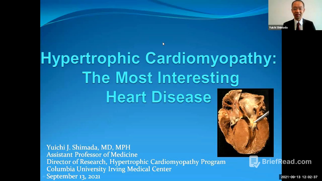
Dr. Shimada introduces hypertrophic cardiomyopathy (HCM) by presenting a case of a young basketball player who died suddenly, with an autopsy revealing HCM. HCM is highlighted as the most common cause of sudden cardiac death in young athletes in the US, distinguishing it from Europe where other conditions like arrhythmogenic RV dysplasia are more prevalent. The gross pathology of HCM shows a thick left ventricle and a narrow left ventricular outflow tract. Microscopic findings reveal myocyte disarray and interstitial fibrosis. Over 1500 mutations in more than a dozen genes are responsible for HCM, but genetic testing has limitations, including challenges in interpreting the significance of each mutation, a high percentage of patients with no identifiable mutations, and weak genetic-clinical correlations.
Diagnosis of Hypertrophic Cardiomyopathy [9:01]
The diagnosis of HCM is characterized by left ventricular hypertrophy without other conditions that can cause the same degree of hypertrophy. The prevalence is 0.2 to 0.5%, affecting one in 200 to 500 people. HCM is often familial with an autosomal dominant pattern of inheritance, leading to diastolic dysfunction, heart failure, ischemia, arrhythmias, and mitral valve abnormalities. Obstructive HCM (HOCM) involves systolic anterior motion (SAM) of the mitral valve, causing LVOT obstruction and mitral regurgitation. Symptoms include dyspnea, angina, palpitations, lightheadedness, dizziness, pre-syncope, or syncope. Physical exam findings may include a brisk carotid upstroke, forceful LV impulse, S4, and a murmur that increases with the Valsalva maneuver.
EKG Findings in HCM [15:22]
EKG findings in HCM can vary widely, showing left ventricular hypertrophy with strain patterns, pseudo-infarct patterns, and giant negative T-waves, particularly in apical HCM. Some patients may exhibit ST elevation, mimicking myocardial infarction. It's advised that patients carry a copy of their EKG to avoid misdiagnosis in emergency situations. In rare cases, patients with HCM may have completely normal EKGs.
Diagnostic Modalities and HCM Mimickers [22:40]
Echocardiography is the most important modality to diagnose HCM, revealing a thick intraventricular septum and systolic anterior motion of the mitral valve. Other conditions that can cause LVH and mimic HCM include pressure overload due to hypertension or aortic stenosis, athlete's heart, Fabry disease, cardiac amyloidosis, and cardiac sarcoidosis. Distinguishing between athlete's heart and HCM can be challenging, as highlighted by the case of Eddie Curry, whose diagnosis affected his career.
Classification and Hemodynamics of HCM [28:03]
HCM is classified into obstructive and non-obstructive categories based on the presence of LVOT obstruction. Obstructive HCM is defined by an LVOT gradient above 30 mmHg at rest or with provocation. Provocation can be achieved through exercise or Valsalva maneuver. Cardiac MRI can be helpful in challenging cases, accurately measuring LV wall thickness and assessing for LV fibrosis using late gadolinium enhancement. Genetic testing can rule in HCM if a pathogenic mutation is found but does not rule it out if negative.
Management of Hypertrophic Cardiomyopathy [34:33]
The management of HCM involves activity restrictions, family screening, prevention of sudden cardiac death, and control of symptoms. Patients are advised against strenuous exercise, competitive sports, and isometric exercise. Family screening strategies include phenotypic screening with echocardiogram and electrocardiogram every three to five years in adults and every one to three years in children and adolescents, as well as genotypic screening. Genetic testing results may influence insurability, so this should be discussed with family members before testing.
Prevention of Sudden Cardiac Death with ICDs [38:35]
ICDs can prevent sudden cardiac death due to ventricular tachycardia or fibrillation, but they are associated with complications. ICD implantation is recommended for patients with prior cardiac arrest or sustained VT. For primary prevention, five major risk factors are considered: family history of sudden cardiac death, unexplained syncope, marked LV hypertrophy, apical aneurysm, and LV ejection fraction equal to or lower than 50%. If none of these are present, extensive late gadolinium enhancement on cardiac MRI and non-sustained ventricular tachycardia on Holter monitoring are considered.
Symptom Control in Obstructive and Non-Obstructive HCM [44:16]
Symptom control in obstructive HCM targets the hypertrophied septum using negative inotropic agents like beta-blockers and calcium channel blockers. Disopyramide can also be used due to its negative inotropic and vasoconstrictive effects. Invasive options include septal myectomy and alcohol septal ablation. The most common complication after alcohol septal ablation is conduction defects requiring a permanent pacemaker. In non-obstructive HCM, therapeutic options are limited to beta-blockers and calcium channel blockers, as disopyramide and invasive procedures are not indicated.


