
TLDR;
This video provides a comprehensive overview of the anatomy of the thorax, including the ribcage, vertebrae, sternum, and related structures. It describes the types of ribs, their connections, and key landmarks within the thoracic cavity, as well as the relationship between the lungs and pleura. Additionally, it touches on potential issues that can arise in the thoracic cavity, such as pneumothorax.
- The thorax consists of the rib cage, sternum, and vertebrae.
- Ribs are categorized into true, false, and floating ribs, each with specific characteristics and attachments.
Introduction to Thoracic Anatomy [0:05]
The video begins by introducing the thoracic anatomy, highlighting the rib cage from an anterior view and detailing the 12 thoracic vertebrae, from T1 to T12. The lumbar vertebra 1 is also identified. The ribs connect to both the vertebrae and the sternum, allowing expansion during respiration. The sternum consists of three parts: the manubrium, the body, and the xiphoid process.
Sternal Angle and Structures [1:00]
The sternal angle, or the angle of Louis, is marked between the manubrium and the body of the sternum. This landmark is significant since various structures, including the second rib articulation and the aortic arch, align with it. A horizontal cut along this angle can reveal several important anatomical features such as the bifurcation of the pulmonary trunk and trachea, as well as the ligamentum arteriosum.
Types of Ribs [2:46]
Ribs are classified into true ribs (1-7), false ribs (8-10), and floating ribs (11-12). True ribs attach directly to the sternum, while false ribs connect to the sternum indirectly. Floating ribs do not attach to the sternum at all. Additionally, ribs can be categorized as typical (ribs 3-9 with similar features) and atypical (ribs 1, 2, and 10-12, which differ).
Vertebrae Overview [4:05]
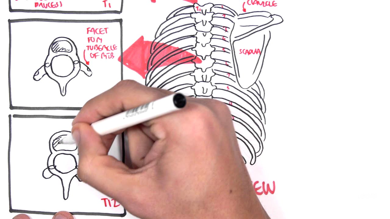
The video provides a posterior view of the thoracic cavity, detailing the thoracic vertebrae structure. T1's body is noted, along with features such as the vertebral foramen where the spinal cord passes and the spinous process. T6 and T12 vertebrae are discussed, particularly the facets where ribs articulate, a defining characteristic of thoracic vertebrae.
Thoracic Cavity Organ Layout [5:30]
Examining the thoracic cavity further, the diaphragm's position relative to the fifth rib is explained. Several important landmarks, including midclavicular, midscapular, and midaxillary lines, are discussed for locating organs within the thorax—especially the lungs and pleura. The pleura's role as a membrane enveloping the lungs is also detailed.
Margins of the Lung and Pleura [6:28]
The video outlines the lung margins based on important anatomical lines, explaining how they differ slightly between the left and right sides due to the heart's location. For example, the midclavicular line for the left lung begins at the fourth rib instead of the sixth, which is the case for the right lung. The margins surrounding the pleura are also addressed, showcasing how they extend further down than the lung itself.
Pulmonary Issues: Pneumothorax [9:09]
The risks associated with the thoracic cavity, specifically pneumothorax—a collapsed lung—are discussed. Pneumothorax can occur when external trauma punctures the parietal pleura, allowing air into the pleural cavity and causing the lung to deflate. Additionally, it can happen due to damage to the visceral pleura in certain lung diseases, like COPD.
Conclusion [10:48]
The video wraps up with a brief recap of the thoracic anatomy and its significance, emphasizing the interrelationships between the structures covered.