TLDR;
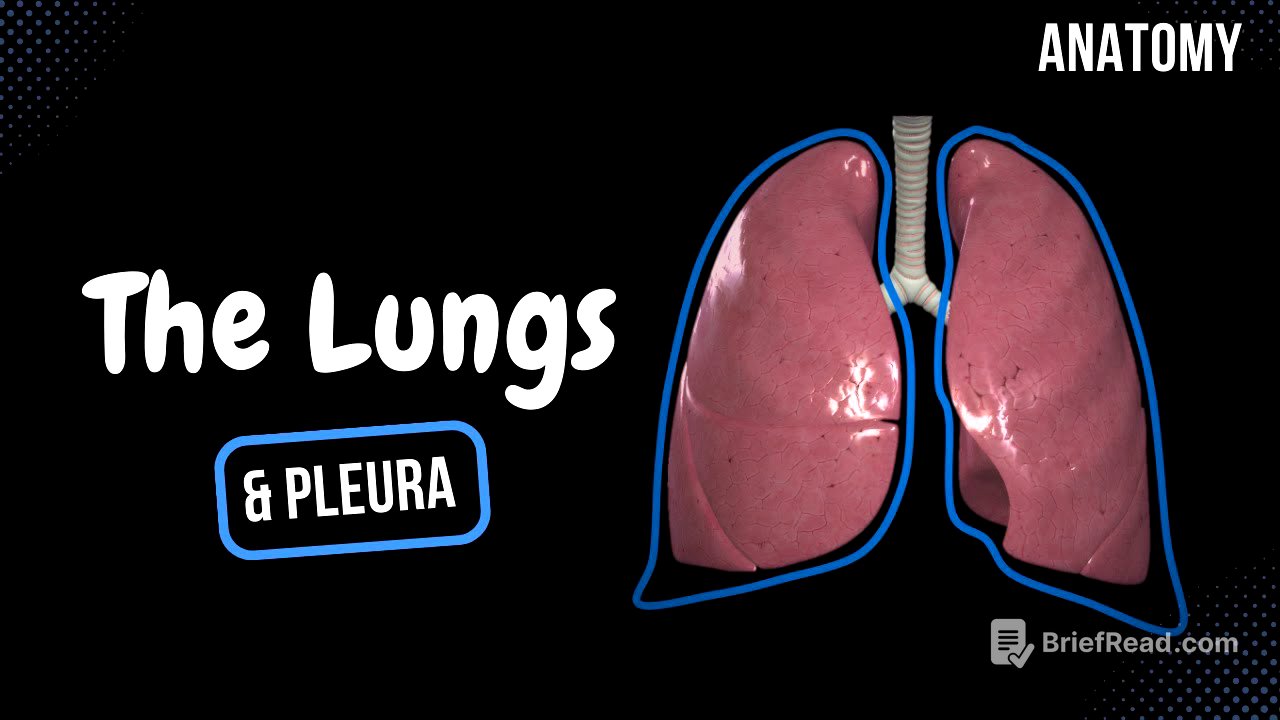
Alright, so this video is all about the anatomy of the lungs and pleura. We're gonna cover lung function, parts, surfaces, margins, lobes, and segments. Then we'll move on to the pleura and finish up with the mediastinum. Key takeaways include understanding the functional units of the lungs (alveoli), the different surfaces and margins, the division into lobes and segments for surgical purposes, the layers of the pleura, and the compartments of the mediastinum.
- Lungs are the primary organ for respiration, aided by muscles of inspiration and expiration.
- Lungs are divided into lobes and segments, each with its own blood and air supply.
- Pleura has visceral and parietal layers, forming the pulmonary ligament.
- Mediastinum is the space between the lungs, divided into superior, anterior, middle, and posterior regions.
Lung Function [0:54]
The lungs are the main organs for breathing, but they don't work alone. Muscles of inspiration, like the diaphragm and external intercostals, help expand the chest cavity so air can come in. There are also muscles of expiration, like the internal intercostals and abdominal muscles, which help push air out, even though this usually happens passively. The functional unit of the lungs are the alveolar sacs, which are highly vascularized. This allows oxygen to enter the bloodstream and carbon dioxide to be removed through diffusion.
Parts and Surfaces of the Lungs [2:04]
The apex of the lung is the cone-shaped top part, and the base is the bottom part. There are four surfaces: the costal surface (facing the ribs), the diaphragmatic surface (facing the diaphragm), the mediastinal surface (facing the space between the lungs), and the interlobar surface (between the lobes). The mediastinal surface is important because it contains the hilum of the lung.
Hilum of the Lung [3:01]
The hilum is like an entrance or hallway into the lung, bordered by the pulmonary ligament. All the structures that enter and exit the lung are called the root of the lung. In the right lung, the bronchus is the highest structure entering the lung, followed by the pulmonary arteries and then the pulmonary veins. In the left lung, the pulmonary artery is the highest, then the bronchus, and then the pulmonary veins. Remember "Bright is Right" (Bronchus is highest in the Right lung).
Parts and Surfaces of the Lungs (revisited) [4:17]
The interlobar surface is a small surface that indicates the separation between the lobes.
Margins of the Lungs [4:33]
Each lung has two margins: the inferior margin, which is thin and sharp and separates the costal and mediastinal surfaces from the diaphragmatic surface, and the anterior margin, which is vertical. The anterior margin is different in each lung. The right lung's anterior margin is almost vertical, but the left lung has a cardiac notch because the heart's apex points to the left. This notch is bordered by a thin piece of lung tissue called the lingula.
Pulmonary Lobes [5:22]
The lungs are divided into lobes by fissures. Both lungs have an oblique fissure, but the right lung also has a horizontal fissure. These fissures divide the lungs into superior, inferior, and middle lobes. Only the right lung has a middle lobe. Each lobe is further divided into segments, which is important for surgical removal of lung tissue (like tumors) without harming other segments in the same lobe. Each lobe has its own blood and air supply.
Segments of Right Lung [6:31]
The right lung has three lobes (superior, middle, and inferior) and a total of 10 segments. The superior lobe has the apical, posterior, and anterior segments. The middle lobe has the lateral and medial segments. The inferior lobe has the superior, basal medial, basal anterior, basal lateral, and basal posterior segments.
Segments of Left Lung [7:27]
The left lung has two lobes (superior and inferior) and usually 8 segments, though some people might have 9 due to variations. The superior lobe has the apicoposterior, anterior, superior lingular, and inferior lingular segments. The inferior lobe has the superior, basal anterior, basal lateral, and basal posterior segments. Sometimes, the basal anterior segment is divided into basal anterior and basal medial segments.
Pleura of the Lungs [8:20]
The pleura are the coverings of the lungs. The innermost layer is the visceral pleura, which is directly on the lungs. The outer layer is the parietal pleura, which is in contact with surrounding structures. The parietal pleura is divided into costal, diaphragmatic, mediastinal parts, and the pleural cupula (covering the apex of the lung). The visceral and parietal pleura meet at the hilum of the lung, forming a double layer called the pulmonary ligament. Between the two layers is the pleural cavity, filled with serous fluid that acts as a lubricant.
Mediastinum [11:01]
The mediastinum is the space between the lungs filled with structures. It's divided into the superior mediastinum and the inferior mediastinum. The inferior mediastinum is further divided into the anterior, middle, and posterior mediastinum. Each region contains specific organs, arteries, and nerves. The superior mediastinum contains the esophagus, trachea, thymus, blood vessels, and nerves. The anterior mediastinum contains blood vessels and part of the pericardium. The middle mediastinum contains the heart and pericardium. The posterior mediastinum contains blood vessels, nerves, and the esophagus.


