TLDR;
This module provides an overview of the muscular system, focusing on its functions, muscle types, and the structure of skeletal muscles. It also covers major muscle groups involved in movement at different joints, offering insights into muscle nomenclature to aid understanding and recall.
- Functions of muscles include movement production, posture maintenance, joint stability, heat production, and organ protection.
- Muscles are classified as voluntary/involuntary, striated/smooth, and somatic/visceral.
- Skeletal muscles are crucial for human movement and possess properties like irritability, contractility, extensibility, and elasticity.
Introduction to the Muscular System [0:00]
The muscular system is vital for understanding human movement and optimising physical performance. The human body contains over 600 muscles, each with specific functions. The primary function of muscles is to produce movement by generating tension that is transferred to bones. Skeletal muscles are under conscious control, enabling deliberate movements like walking and lifting. Smooth muscles in organs like the digestive tract and cardiac muscles in the heart operate involuntarily, facilitating processes such as digestion and blood circulation.
Functions of Muscles [1:18]
Muscles play several key roles in the body. They maintain posture by working against gravity and supporting the spine. They contribute to joint stability, reducing the risk of injuries. Muscle contraction generates heat, which helps maintain body temperature and contributes to homeostasis. Muscles also protect vital organs by providing support and acting as a barrier against external forces. Additional functions include facilitating breathing through muscles like the diaphragm, regulating blood flow via smooth muscles in blood vessels, and aiding in swallowing with muscles in the throat and oesophagus.
Classification of Muscles [4:58]
Muscles are classified based on distinct characteristics. They can be voluntary (consciously controlled, like skeletal muscles) or involuntary (not consciously controlled, like cardiac and smooth muscles). Another classification is based on whether they are striated (striped appearance under a microscope, like skeletal and cardiac muscles) or smooth (unstriated). Muscles are also categorised as somatic (present in body walls and limbs) or visceral (making up hollow organs like the digestive tract).
Muscle Types: Skeletal, Cardiac, and Smooth [7:42]
Skeletal muscles are attached to the skeleton or fascia, producing movement through shortening and relaxation. They also maintain posture. Cardiac muscles, found in the heart and adjacent great vessels, are strong, quick to respond, and provide continuous rhythmic contractions for pumping blood. Smooth muscles are present in the walls of hollow viscera, blood vessels, and skin follicles. They are weaker, have slower contractions, and help propel substances and restrict fluid flow in the body.
Characteristics of Skeletal Muscles [9:37]
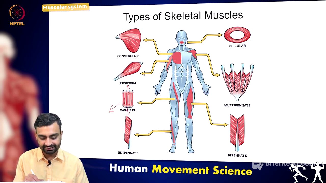
Skeletal muscles are resilient and can be stretched and shortened at high speeds without significant damage. Key properties include irritability (ability to respond to stimulation), contractility (ability to shorten with sufficient stimulation), extensibility (ability to lengthen beyond resting length), and elasticity (ability to return to resting length after stretch). Skeletal muscles are categorised by fibre arrangement, including parallel, convergent, fusiform, circular, unipennate, bipennate, and multipennate types, each suited for different movements and force generation.
Structure of Skeletal Muscles [15:12]
Skeletal muscles attach to bones via tendons. The entire muscle bundle is wrapped in epimysium, which contains fascicles wrapped in perimysium. Fascicles consist of muscle fibres, each wrapped in endomysium. Muscle fibres are made up of myofibrils, which contain repetitive units called sarcomeres. Sarcomeres are composed of actin and myosin filaments; their overlap varies during contraction, relaxation, and extension.
Major Muscle Groups in the Upper Limb [16:32]
Major muscle groups in the upper body include the trapezius, deltoid, pectoral muscles, biceps, triceps, latissimus dorsi, abdominal muscles, and teres group. Understanding muscle nomenclature aids in remembering names and functions. For example, "abductor digiti minimi" consists of "ab" (away from), "ductor" (to move), "digiti" (digit), and "minimi" (tiny), indicating a muscle that moves the little finger or toe away. Common root words include "ad" (toward), "sub" (under), "longissimus" (longest), "maximus" (large), and "externus" (outside).
Muscle Groups at the Shoulder Joint [20:31]
The major muscle groups at the shoulder joint are categorised into four groups: extrinsic (superficial trapezius and latissimus dorsi, and deep levator scapulae, rhomboid major and minor), intrinsic (deltoid and teres major), rotator cuff (supraspinatus, infraspinatus, subscapularis, and teres minor), and pectoral (pectoralis major and minor, and serratus anterior). These muscles facilitate movements such as extension (posterior deltoid, latissimus dorsi, teres major), flexion (biceps brachii, pectoralis major, anterior deltoid, coracobrachialis), abduction (supraspinatus, deltoid, trapezius, serratus anterior), adduction (pectoralis major, latissimus dorsi, teres major), medial rotation (subscapularis, pectoralis major, latissimus dorsi, teres major, anterior deltoid), and lateral rotation (infraspinatus, teres minor).
Muscle Groups at the Elbow Joint [24:34]
The elbow joint has four major muscle groups, subdivided into upper arm (anterior compartment: biceps brachii, coracobrachialis, brachialis; posterior compartment: triceps brachii) and forearm (anterior compartment: pronator teres, pronator quadratus; posterior compartment: anconeus, brachioradialis, supinator). These muscles produce movements such as extension (triceps brachii, anconeus), flexion (brachialis, biceps brachii, brachioradialis), pronation (pronator quadratus, pronator teres), and supination (supinator, biceps brachii).
Muscle Groups at the Wrist Joint [26:53]
Most muscles controlling wrist movement are located in the forearm, with some contribution from hand muscles. Movements include flexion (flexor carpi ulnaris, flexor carpi radialis, palmaris longus, flexor digitorum superficialis), extension (extensor carpi radialis longus and brevis, extensor carpi ulnaris, extensor digitorum), radial deviation (flexor carpi radialis, extensor carpi radialis), and ulnar deviation (flexor carpi ulnaris, extensor carpi ulnaris).
Major Muscle Groups in the Lower Limb [29:29]
The lower limb's major muscle groups are located at the hip, knee, and ankle joints. The hip joint includes the gluteal group (gluteus maximus, medius, minimus, tensor fascia latae), adductor group (adductor brevis, longus, magnus, pectineus, gracilis), iliopsoas group (iliacus, psoas major), and lateral rotator group (external and internal obturator, piriformis, superior and inferior gemelli, quadratus femoris). Movements at the hip include extension (gluteus maximus, adductor magnus, biceps femoris), flexion (gracilis, psoas major, iliacus, pectineus), abduction (gluteus medius and minimus, obturator externus, gemelli, sartorius), adduction (adductor group), and rotation (lateral rotator group, biceps femoris, sartorius, gluteus medius and minimus).
Muscle Groups at the Knee Joint [32:41]
The knee joint has three major muscle groups: the hamstring group (biceps femoris, semitendinosus, semimembranosus), the quadriceps femoris (vastus lateralis, intermedius, medialis, rectus femoris), and the popliteus. Movements at the knee include extension (sartorius, quadriceps femoris), flexion (biceps femoris, semitendinosus, semimembranosus), and rotation (biceps femoris, semitendinosus, semimembranosus, gracilis, sartorius). The popliteus muscle facilitates knee flexion by unlocking the fully extended knee joint.
Muscle Groups at the Ankle Joint [34:44]
Ankle joint movement is primarily controlled by muscles in the lower leg. The major muscle groups include the anterior compartment (tibialis anterior, extensor digitorum longus, extensor hallucis longus), posterior compartment (superficial: gastrocnemius, plantaris, soleus; deep: tibialis posterior), and lateral compartment (fibularis longus and brevis). Movements at the ankle include eversion (fibularis brevis and longus), inversion (tibialis posterior and anterior), dorsiflexion (tibialis anterior, extensor hallucis longus, extensor digitorum longus), and plantar flexion (gastrocnemius, plantaris, soleus, fibularis longus).